Explore
 Validate
 Learn
Learn Western blot
 ELISA
ELISAAntibody data
- Antibody Data
- Antigen structure
- References [0]
- Comments [0]
- Validations
- Western blot [2]
- Immunohistochemistry [1]
Submit
Validation data
Reference
Comment
Report error
- Product number
- NBP1-77822-0.1mg - Provider product page

- Provider
- Novus Biologicals
- Product name
- Rabbit Polyclonal Nrf1 Antibody
- Antibody type
- Polyclonal
- Description
- Protein A purified. This protein A purified antibody is directed against mouse NRF1. BLAST analysis was used to suggest reactivity with this protein from mouse, human, chimpanzee, dog.
- Reactivity
- Mouse
- Host
- Rabbit
- Isotype
- IgG
- Vial size
- 0.1 mg
- Concentration
- 2.2 mg/ml
- Storage
- Store at -20C. Avoid freeze-thaw cycles.
No comments: Submit comment
Supportive validation
- Submitted by
- Novus Biologicals (provider)
- Main image
- Experimental details
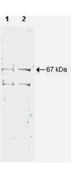
- Western Blot: Nrf1 Antibody [NBP1-77822] - Shows detection of a 67-kDa band corresponding to human NRF1 in a (lane 1) HeLa nuclear extract and (lane 2) whole cell lysate (molecular weight marker not shown). Approx. 10 ug of each lysate was separated by SDS-PAGE and transferred onto nitrocellulose. The blot was incubated with a 1:500 dilution of the antibody at room temperature for 1 h followed by detection using IRDye (TM) 700 labeled Goat-a-Rabbit IgG [H&L] diluted 1:2,500. IRDye (TM) 700

- Submitted by
- Novus Biologicals (provider)
- Main image

- Experimental details
- Western Blot: Nrf1 Antibody [NBP1-77822] - Lane: NFR1-HIS recombinant protein. Load: 50ug per lane. Primary antibody - 1: NRF1 antibody at 1:1,000 overnight at 4C. Primary antibody - 2: 6xHIS Epitope tag antibody at 1:1,000 overnight at 4C. Secondary antibody: Peroxidase rabbit secondary antibody at 1:40,000 for 30 min at RT. Blocking: MB-070 for 30 min at RT. Predicted/observed size: 67 kDa, 67 kDa for NRF1-His tagged. Other band(s): None.

Supportive validation
- Submitted by
- Novus Biologicals (provider)
- Main image

- Experimental details
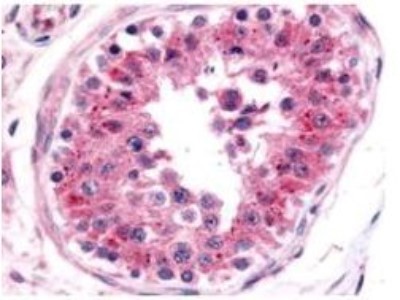
- Immunohistochemistry: Nrf1 Antibody [NBP1-77822] - Used at a 5 ug/ml to detect NRF1 in a variety of tissues. This image shows NRF1 staining of human testis. The antibody shows nuclear staining in lymphocytes. Tissue was formalin-fixed and paraffin embedded.