Explore
 Validate
 Learn
Learn Western blot
 Immunocytochemistry
Antibody data
- Antibody Data
- Antigen structure
- References [1]
- Comments [0]
- Validations
- Immunocytochemistry [5]
- Immunohistochemistry [4]
- Flow cytometry [2]
- Other assay [1]
Submit
Validation data
Reference
Comment
Report error
- Product number
- MA5-32782 - Provider product page

- Provider
- Invitrogen Antibodies
- Product name
- NRF1 Recombinant Rabbit Monoclonal Antibody (JM89-63)
- Antibody type
- Monoclonal
- Antigen
- Synthetic peptide
- Description
- Recombinant rabbit monoclonal antibodies are produced using in vitro expression systems. The expression systems are developed by cloning in the specific antibody DNA sequences from immunoreactive rabbits. Then, individual clones are screened to select the best candidates for production. The advantages of using recombinant rabbit monoclonal antibodies include: better specificity and sensitivity, lot-to-lot consistency, animal origin-free formulations, and broader immunoreactivity to diverse targets due to larger rabbit immune repertoire.
- Reactivity
- Human, Mouse, Rat
- Host
- Rabbit
- Isotype
- IgG
- Antibody clone number
- JM89-63
- Vial size
- 100 μL
- Concentration
- 1 mg/mL
- Storage
- Store at 4°C short term. For long term storage, store at -20°C, avoiding freeze/thaw cycles.
Submitted references Alcalase Potato Protein Hydrolysate-PPH902 Enhances Myogenic Differentiation and Enhances Skeletal Muscle Protein Synthesis under High Glucose Condition in C2C12 Cells.
Chen YJ, Chang CF, Angayarkanni J, Lin WT
Molecules (Basel, Switzerland) 2021 Oct 30;26(21)
Molecules (Basel, Switzerland) 2021 Oct 30;26(21)
No comments: Submit comment
Supportive validation
- Submitted by
- Invitrogen Antibodies (provider)
- Main image
- Experimental details
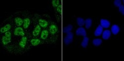
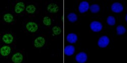
- Immunocytochemical analysis of NRF1 in Hela cells using a NRF1 Monoclonal antibody (Product # MA5-32782) as seen in green. The nuclear counter stain is DAPI (blue). Cells were fixed in paraformaldehyde, permeabilised with 0.25% Triton X100/PBS.
- Submitted by
- Invitrogen Antibodies (provider)
- Main image

- Experimental details
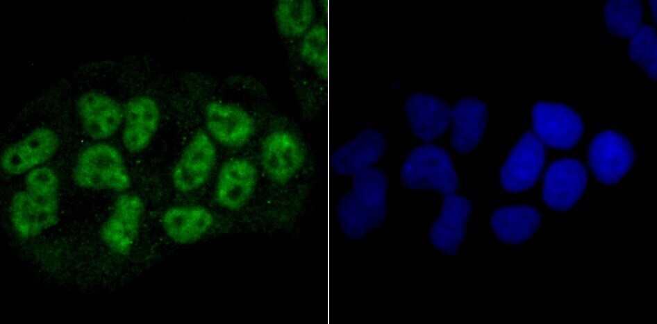
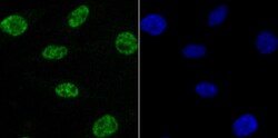
- Immunocytochemical analysis of NRF1 in MCF-7 cells using a NRF1 Monoclonal antibody (Product # MA5-32782) as seen in green. The nuclear counter stain is DAPI (blue). Cells were fixed in paraformaldehyde, permeabilised with 0.25% Triton X100/PBS.
- Submitted by
- Invitrogen Antibodies (provider)
- Main image

- Experimental details
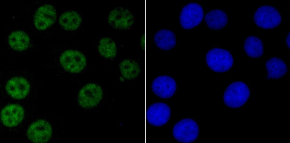
- Immunocytochemical analysis of NRF1 in SH-SY5Y cells using a NRF1 Monoclonal antibody (Product # MA5-32782) as seen in green. The nuclear counter stain is DAPI (blue). Cells were fixed in paraformaldehyde, permeabilised with 0.25% Triton X100/PBS.

- Submitted by
- Invitrogen Antibodies (provider)
- Main image
- Experimental details
- Immunocytochemical analysis of NRF1 in MCF-7 cells using a NRF1 Monoclonal antibody (Product # MA5-32782) as seen in green. The nuclear counter stain is DAPI (blue). Cells were fixed in paraformaldehyde, permeabilised with 0.25% Triton X100/PBS.

- Submitted by
- Invitrogen Antibodies (provider)
- Main image
- Experimental details
- Immunocytochemical analysis of NRF1 in SH-SY5Y cells using a NRF1 Monoclonal antibody (Product # MA5-32782) as seen in green. The nuclear counter stain is DAPI (blue). Cells were fixed in paraformaldehyde, permeabilised with 0.25% Triton X100/PBS.

Supportive validation
- Submitted by
- Invitrogen Antibodies (provider)
- Main image
- Experimental details
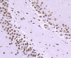
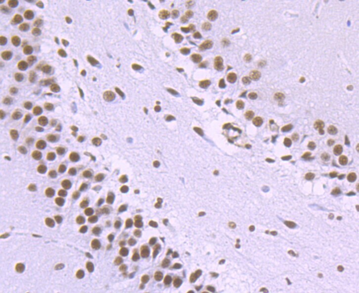
- Immunohistochemical analysis of NRF1 of paraffin-embedded rat brain tissue using a NRF1 Monoclonal antibody (Product #MA5-32782). Counter stained with hematoxylin.
- Submitted by
- Invitrogen Antibodies (provider)
- Main image
- Experimental details
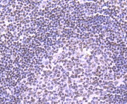
- Immunohistochemical analysis of NRF1 of paraffin-embedded Human tonsil tissue using a NRF1 Monoclonal antibody (Product #MA5-32782). Counter stained with hematoxylin.

- Submitted by
- Invitrogen Antibodies (provider)
- Main image

- Experimental details
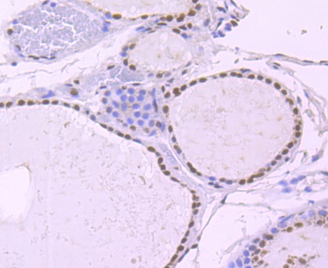
- Immunohistochemical analysis of NRF1 of paraffin-embedded Human thyroid tissue using a NRF1 Monoclonal antibody (Product #MA5-32782). Counter stained with hematoxylin.
- Submitted by
- Invitrogen Antibodies (provider)
- Main image
- Experimental details
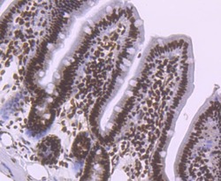
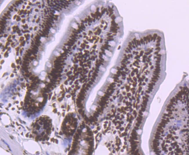
- Immunohistochemical analysis of NRF1 of paraffin-embedded Mouse colon tissue using a NRF1 Monoclonal antibody (Product #MA5-32782). Counter stained with hematoxylin.
Supportive validation
- Submitted by
- Invitrogen Antibodies (provider)
- Main image

- Experimental details
- Flow Cytometric analysis of NRF1 in 293T cells using a NRF1 Monoclonal Antibody (Product # MA5-32782) at a dilution of 1:100, as seen in red compared with an unlabelled control (cells without incubation with primary antibody; black).
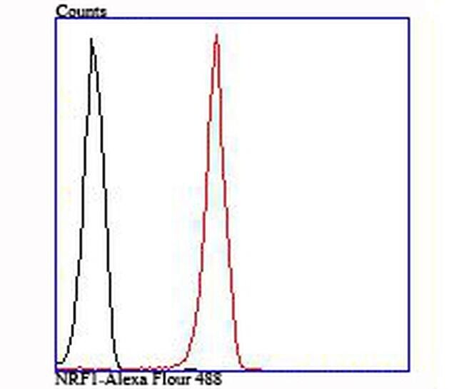
- Submitted by
- Invitrogen Antibodies (provider)
- Main image
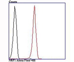
- Experimental details
- Flow Cytometric analysis of NRF1 in 293T cells using a NRF1 Monoclonal Antibody (Product # MA5-32782) at a dilution of 1:100, as seen in red compared with an unlabelled control (cells without incubation with primary antibody; black).

Supportive validation
- Submitted by
- Invitrogen Antibodies (provider)
- Main image
- Experimental details
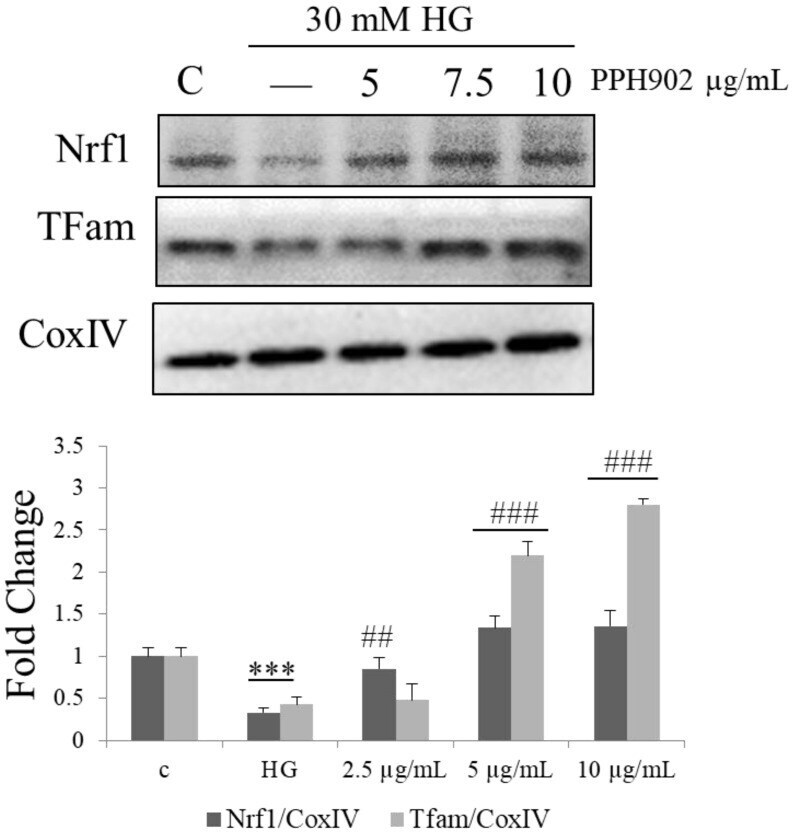
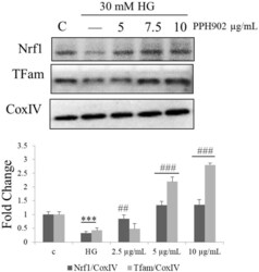
- Figure 9 Effect of PPH902 on the activation of mitochondrial biogenesis: Representative Western blotting shows changes in the levels of NRF1 and TFAM in C2C12 cells. *** p < 0.001 indicates a significant difference with respect to control groups and ## p < 0.01 and ### p < 0.001 indicates a significance difference with respect to the high glucose challenge groups.