Explore
 Validate
 Learn
Learn Western blot
 ELISA
ELISAAntibody data
- Antibody Data
- Antigen structure
- References [0]
- Comments [0]
- Validations
- Western blot [1]
- Immunohistochemistry [1]
Submit
Validation data
Reference
Comment
Report error
- Product number
- AP09128PU-N - Provider product page

- Provider
- Acris Antibodies GmbH
- Proper citation
- Acris Antibodies GmbH Cat#AP09128PU-N, RRID:AB_2035805
- Product name
- anti NRF1 (1-534)
- Antibody type
- Polyclonal
- Antigen
- Recombinant Mouse NRF1 protein corresponding to aa 1-534 of the native protein.
- Reactivity
- Human, Mouse, Rat, Chicken/Avian, Zebrafish
- Host
- Rabbit
- Isotype
- IgG
- Vial size
- 0.5 mg
- Concentration
- 2.3 mg/ml (by UV absorbance at 280 nm)
No comments: Submit comment
Supportive validation
- Submitted by
- Acris Antibodies GmbH (provider)
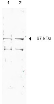
- Main image
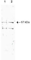
- Experimental details
- Western blot using Protein A Purified AP09128PU-N NRF1 antibody shows detection of a 67-kDa band corresponding to Human NRF1 in a (Lane 1) HeLa nuclear extract and (Lane 2) whole cell lysate (molecular weight marker not shown). ~10 µg of each lysate was separated by SDS-PAGE and transferred onto nitrocellulose. The blot was incubated with a 1/500 dilution of the antibody at RT for 1 h followed by detection using IRDyeâ¢700 labeled Goat anti-Rabbit IgG [H&L] diluted 1/2,500. IRDyeâ¢700 fluorescence image was captured using the Odyssey® Infrared Imaging System developed by LI-COR. IRDye is a trademark of LI-COR, Inc. Other detection systems will yield similar results.
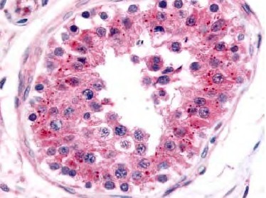
Supportive validation
- Submitted by
- Acris Antibodies GmbH (provider)
- Main image

- Experimental details
- Immunohistochemistry. AP09128PU-N NRF1 antibody staining of Formalin-Fixed, Paraffin Embedded Human testis. This Affinity purified antibody was used at 5 µg/ml to detect NRF1 in a variety of tissues. It shows nuclear staining in lymphocytes.Personal Communication, Tina Roush, LifeSpanBiosciences, Seattle, WA.