 Explore
 Validate
 Learn
Learn Western blot
Antibody data
- Antibody Data
- Antigen structure
- References [0]
- Comments [0]
- Validations
- Western blot [2]
- Immunocytochemistry [1]
- Immunohistochemistry [2]
Submit
Validation data
Reference
Comment
Report error
- Product number
- PA5-41820 - Provider product page

- Provider
- Invitrogen Antibodies
- Product name
- IGF2BP1 Polyclonal Antibody
- Antibody type
- Polyclonal
- Antigen
- Synthetic peptide
- Description
- Peptide sequence: PDEQIAQGPE NGRRGGFGSR GQPRQGSPVA AGAPAKQQQV DIPLRLLVPT Sequence homology: Cow: 93%; Dog: 100%; Guinea Pig: 93%; Horse: 93%; Human: 100%; Mouse: 93%; Pig: 100%; Rabbit: 100%; Rat: 100%; Sheep: 93%
- Reactivity
- Human, Rat
- Host
- Rabbit
- Isotype
- IgG
- Vial size
- 100 µL
- Concentration
- 0.5 mg/mL
- Storage
- -20° C, Avoid Freeze/Thaw Cycles
No comments: Submit comment
Supportive validation
- Submitted by
- Invitrogen Antibodies (provider)
- Main image

- Experimental details
- Western blot analysis of human HepG2 cell lysate using an anti-IGF2BP1 polyclonal antibody (Product # PA5-41820).

- Submitted by
- Invitrogen Antibodies (provider)
- Main image
- Experimental details
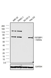
- Western blot was performed using Anti-IGF2BP1 Polyclonal Antibody (Product # PA5-41820) and 62 kDa band along with a higher molecular weight at ~ 120 kDa (probable glycosylated form) corresponding to IGF2BP1 was observed across all the cell lines tested except SK-OV-3. Whole cell extracts (30 µg lysate) of IMR-32 (Lane 1), PANC-1 (Lane 2), SK-OV-3 (Lane 3) and Hep G2 (Lane 4) were electrophoresed using NuPAGE™ 4-12% Bis-Tris Protein Gel (Product # NP0322BOX). Resolved proteins were then transferred onto a nitrocellulose membrane (Product # IB23001) by iBlot® 2 Dry Blotting System (Product # IB21001). The blot was probed with the primary antibody (1 µg/ml) and detected by chemiluminescence with Goat anti-Rabbit IgG (H+L) Superclonal™ Recombinant Secondary Antibody, HRP (Product # A27036, 1:4000 dilution) using the iBright FL 1000 (Product # A32752). Chemiluminescent detection was performed using Novex® ECL Chemiluminescent Substrate Reagent Kit (Product # WP20005).
Supportive validation
- Submitted by
- Invitrogen Antibodies (provider)
- Main image
- Experimental details
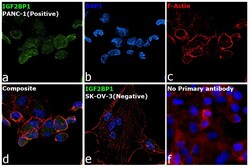
- Immunofluorescence analysis of IGF2BP1 was performed using 70% confluent log phase PANC-1 and SK-OV-3 cells. The cells were fixed with 4% Paraformaldehyde for 10 minutes, permeabilized with 0.1% Triton™ X-100 for 10 minutes, and blocked with 2% BSA for 1 hour at room temperature. The cells were labeled with IGF2BP1 Polyclonal Antibody (Product # PA5-41820) at 1:100 dilution in 0.1% BSA, incubated at 4 degree celsius overnight and then labeled with Goat anti-rabbit IgG (H+L) Superclonal™ Secondary Antibody, Alexa Fluor® 488 conjugate (Product # A27034, 1:2000 dilution) for 45 minutes at room temperature (Panel a: Green). Nuclei (Panel b: Blue) were stained with SlowFade® Gold Antifade Mountant with DAPI (Product # S36938). F-actin (Panel c: Red) was stained with Rhodamine Phalloidin (Product # R415, 1:300). Panel d represents the merged image showing nuclear and cytoplasmic localization. Panel e represents SK-OV-3 cells having no expression of IG2BP1. Panel f represents control cells with no primary antibody to assess background. The images were captured at 60X magnification.

Supportive validation
- Submitted by
- Invitrogen Antibodies (provider)
- Main image

- Experimental details
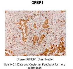
- Immunohistochemistry analysis of human lung adenocarcinoma tissue using an anti-IGF2BP1 polyclonal antibody (Product # PA5-41820).

- Submitted by
- Invitrogen Antibodies (provider)
- Main image
- Experimental details
- Immunohistochemistry analysis of human lung adenocarcinoma cells using an anti-IGF2BP1 polyclonal antibody (Product # PA5-41820). Primary Antibody Dilution:1:300; Secondary Antibody: Anti-rabbit-linker, Fbex-HRP.
