Explore
 Validate
 Learn
Learn Western blot
 Immunocytochemistry
 Gel shift
Antibody data
- Antibody Data
- Antigen structure
- References [1]
- Comments [0]
- Validations
- Immunocytochemistry [2]
- Other assay [1]
Submit
Validation data
Reference
Comment
Report error
- Product number
- 712138 - Provider product page

- Provider
- Invitrogen Antibodies
- Product name
- IGF2BP1 Recombinant Superclonal™ Antibody (18HCLC)
- Antibody type
- Other
- Antigen
- Synthetic peptide
- Description
- This antibody is predicted to react with Monkey, Dog, Cat. Recombinant rabbit Superclonal™ antibodies are unique offerings from Thermo Fisher Scientific. They are comprised of a selection of multiple different recombinant monoclonal antibodies, providing the best of both worlds - the sensitivity of polyclonal antibodies with the specificity of monoclonal antibodies - all delivered with the consistency only found in a recombinant antibody. While functionally the same as a polyclonal antibody - recognizing multiple epitope sites on the target and producing higher detection sensitivity for low abundance targets - a recombinant rabbit Superclonal™ antibody has a known mixture of light and heavy chains. The exact population can be produced in every lot, circumventing the biological variability typically associated with polyclonal antibody production. Note: Formerly called “Recombinant polyclonal antibody”, this product is now rebranded as “Recombinant Superclonal™ antibody”. The physical product and the performance remain unchanged.
- Reactivity
- Human, Mouse
- Host
- Rabbit
- Isotype
- IgG
- Antibody clone number
- 18HCLC
- Vial size
- 100 μg
- Concentration
- 0.5 mg/mL
- Storage
- Store at 4°C short term. For long term storage, store at -20°C, avoiding freeze/thaw cycles.
Submitted references Testicular orphan receptor 4 (TR4) promotes papillary thyroid cancer invasion via activating circ-FNLA/miR-149-5p/MMP9 signaling.
Ouyang X, Feng L, Yao L, Xiao Y, Hu X, Zhang G, Liu G, Wang Z
Molecular therapy. Nucleic acids 2021 Jun 4;24:755-767
Molecular therapy. Nucleic acids 2021 Jun 4;24:755-767
No comments: Submit comment
Supportive validation
- Submitted by
- Invitrogen Antibodies (provider)
- Main image

- Experimental details
- For immunofluorescence analysis,HepG2 cells were fixed and permiabilized detection of endogenous IGF2BP1 using Anti-IGF2BP1 Recombinant Rabbit Polyclonal Antibody (Product # 712138, 1:100) and labeled with Goat anti-Rabbit IgG (H+L) Superclonal™ Secondary Antibody, Alexa Fluor® 488 conjugate (Product # A27034, 1:2000). Nuclei (blue) were stained using SlowFade® Gold Antifade Mountant with DAPI (Product # S36938), and Rhodamine Phalloidin (Product # R415, 1:300) was used for cytoskeletal F-actin (red) staining. Panel a-d) clearly demonstares cytoplasmic localization of IGF2BP1 in HepG2 cells . Panel e-h) shows no specific staining in HT29 cells which is IGF2BP1 negative,demonstrating specificity. The images were captured at 60X magnification.
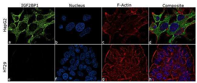
- Submitted by
- Invitrogen Antibodies (provider)
- Main image

- Experimental details
- For immunofluorescence analysis,HepG2 cells were fixed and permiabilized detection of endogenous IGF2BP1 using Anti-IGF2BP1 Recombinant Rabbit Superclonal™ Antibody (Product # 712138, 1:100) and labeled with Goat anti-Rabbit IgG (Heavy Chain) Superclonal™ Secondary Antibody, Alexa Fluor® 488 conjugate (Product # A27034, 1:2000). Nuclei (blue) were stained using SlowFade® Gold Antifade Mountant with DAPI (Product # S36938), and Rhodamine Phalloidin (Product # R415, 1:300) was used for cytoskeletal F-actin (red) staining. Panel a-d) clearly demonstares cytoplasmic localization of IGF2BP1 in HepG2 cells . Panel e-h) shows no specific staining in HT29 cells which is IGF2BP1 negative,demonstrating specificity. The images were captured at 60X magnification.

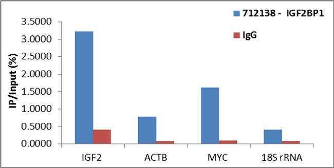
Supportive validation
- Submitted by
- Invitrogen Antibodies (provider)
- Main image
- Experimental details
- Detection of binding of endogenous IGF2BP1 protein to specific RNA using Anti-IGF2BP1 Antibody: RNA Immunoprecipitation (RIP) was performed using Anti-IGF2BP1 Recombinant Rabbit Polyclonal Antibody (Product # 712138, 4 µg) on clarified whole cell lysate from 2 million Hep G2 cells. Normal Rabbit IgG was used as a negative IP control. Immunoprecipitated RNA was purified by RiboPure™ RNA Purification Kit (Product # AM1924) and analyzed by RT-PCR using the Power SYBR® Green RNA-to-CT™ 1-Step Kit (Product # 4389986) with primer pairs over IGF2, MYC, ACTB mRNA (positive) and 18s rRNA (negative). Data is presented as the fraction of immunoprecipitated RNA (%IP) normalized to the total amount of RNA used for immunoprecipitation.