 Explore
 Validate
 Learn
Learn Western blot
Antibody data
- Antibody Data
- Antigen structure
- References [0]
- Comments [0]
- Validations
- Western blot [3]
- Immunocytochemistry [1]
- Immunohistochemistry [1]
- Flow cytometry [1]
Submit
Validation data
Reference
Comment
Report error
- Product number
- PA5-23968 - Provider product page

- Provider
- Invitrogen Antibodies
- Product name
- IGF2BP1 Polyclonal Antibody
- Antibody type
- Polyclonal
- Antigen
- Synthetic peptide
- Description
- This antibody is predicted to react with chicken, mouse, rat and Xenopus and zebrafish based on sequence homology.
- Reactivity
- Human, Mouse
- Host
- Rabbit
- Isotype
- IgG
- Vial size
- 400 µL
- Concentration
- 0.5 mg/mL
- Storage
- Store at 4°C short term. For long term storage, store at -20°C, avoiding freeze/thaw cycles.
No comments: Submit comment
Supportive validation
- Submitted by
- Invitrogen Antibodies (provider)
- Main image
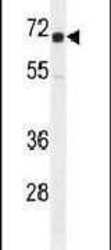
- Experimental details
- Western blot analysis using an IGF2BP1 polyclonal antibody (Product # PA5-23968) in K562 cell lysates (35 µg per lane).

- Submitted by
- Invitrogen Antibodies (provider)
- Main image
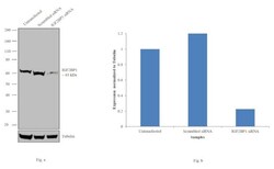
- Experimental details
- Knockdown of IGF2BP1 was achieved by transfecting Hep G2 with IGF2BP1 specific siRNAs (Silencer® select Product # s20916; s20918). Western blot analysis (Fig. a) was performed using whole cell extracts from the IGF2BP1 knockdown cells (lane 3), non-specific scrambled siRNA transfected cells (lane 2) and untransfected cells (lane 1). The blots were probed with IGF2BP1 Polyclonal Antibody (Product # PA5-23968, 1:1000 dilution) and Goat anti-Rabbit IgG (H+L) Superclonal™ Secondary Antibody, HRP conjugate (Product # A27036, 0.25µg/mL, 1:4000 dilution). Densitometric analysis of this western blot is shown in histogram (Fig. b). Decrease in signal upon siRNA mediated knock down confirms that antibody is specific to IGF2BP1.

- Submitted by
- Invitrogen Antibodies (provider)
- Main image
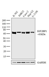
- Experimental details
- Western blot analysis was performed on whole cell extracts (30 µg lysate) of K-562 (Lane 1), IMR32 (Lane 2), Caco-2 (Lane 3), Hep G2 (Lane 4) and U-2 OS (Lane 5). The blot was probed with Anti-IGF2BP1 Polyclonal Antibody (Product # PA5-23968, 1:1000 dilution) and detected by chemiluminescence Goat anti-Rabbit IgG (H+L) Superclonal™ Secondary Antibody, HRP conjugate (Product # A27036, 0.25 µg/mL, 1:4000 dilution). A 63kDa band corresponding to IGF2BP1 was observed across all the cell lines tested.

Supportive validation
- Submitted by
- Invitrogen Antibodies (provider)
- Main image

- Experimental details
- Immunofluorescence analysis of IGF2BP1 was performed using 70% confluent log phase Caco-2 cells. The cells were fixed with 4% paraformaldehyde for 10 minutes, permeabilized with 0.1% Triton™ X-100 for 15 minutes, and blocked with 1% BSA for 1 hour at room temperature. The cells were labeled with anti-IGF2BP1 Polyclonal Antibody (Product # PA5-23968) at 1:100 dilution in 0.1% BSA, incubated at 4 degree Celsius overnight and then labeled with Goat anti-Rabbit IgG (H+L) Superclonal™ Secondary Antibody, Alexa Fluor® 488 conjugate (Product # A27034) at a dilution of 1:2000 for 45 minutes at room temperature (Panel a: green). Nuclei (Panel b: blue) were stained with ProLong™ Diamond Antifade Mountant with DAPI (Product # P36962). F-actin (Panel c: red) was stained with Rhodamine Phalloidin (Product # R415, 1:300). Panel d represents the merged image showing Nuclear and Cytoplasmic localization. Panel e represents control cells with no primary antibody to assess background. The images were captured at 60X magnification

Supportive validation
- Submitted by
- Invitrogen Antibodies (provider)
- Main image

- Experimental details
- Immunohistochemistry analysis in formalin-fixed, paraffin-embedded human prostate carcinoma using an IGF2BP1 polyclonal antibody (Product # PA5-23968), followed by HRP-conjugated secondary antibody and DAB staining.

Supportive validation
- Submitted by
- Invitrogen Antibodies (provider)
- Main image

- Experimental details
- Flow cytometry analysis of K562 cells using an IGF2BP1 polyclonal antibody (Product # PA5-23968) (right) compared to a negative control cell (left) at a dilution of 1:10-50, followed by a FITC-conjugated goat anti-rabbit antibody
