Explore
 Validate
 Learn
Learn Western blot
Antibody data
- Antibody Data
- Antigen structure
- References [1]
- Comments [0]
- Validations
- Western blot [1]
- Immunocytochemistry [2]
Submit
Validation data
Reference
Comment
Report error
- Product number
- AF4050 - Provider product page

- Provider
- R&D Systems
- Product name
- Human/Mouse MYCL1/L-Myc Antibody
- Antibody type
- Polyclonal
- Description
- Immunogen affinity purified. Detects human and mouse MYCL1 / L-Myc in Western blots
- Reactivity
- Human, Mouse
- Host
- Goat
- Conjugate
- Unconjugated
- Antigen sequence
P12524- Isotype
- IgG
- Vial size
- 100 ug
- Concentration
- LYOPH
- Storage
- Use a manual defrost freezer and avoid repeated freeze-thaw cycles. 12 months from date of receipt, -20 to -70 °C as supplied. 1 month, 2 to 8 °C under sterile conditions after reconstitution. 6 months, -20 to -70 °C under sterile conditions after reconstitution.
Submitted references Merkel Cell Polyomavirus Small T Antigen Promotes Pro-Glycolytic Metabolic Perturbations Required for Transformation.
Berrios C, Padi M, Keibler MA, Park DE, Molla V, Cheng J, Lee SM, Stephanopoulos G, Quackenbush J, DeCaprio JA
PLoS pathogens 2016 Nov;12(11):e1006020
PLoS pathogens 2016 Nov;12(11):e1006020
No comments: Submit comment
Supportive validation
- Submitted by
- R&D Systems (provider)
- Main image
- Experimental details
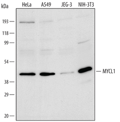
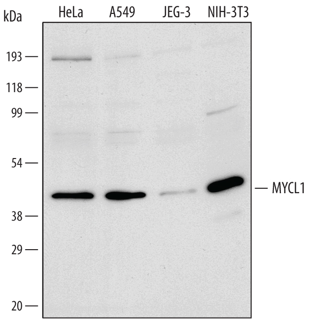
- Detection of Human/Mouse MYCL1/ L-Myc by Western Blot. Western blot shows nuclear extracts of HeLa human cervical epithelial carcinoma cell line, A549 human lung carcinoma cell line, JEG-3 human epithelial choriocarcinoma cell line, and NIH-3T3 mouse embryonic fibroblast cell line. PVDF membrane was probed with 1 µg/mL of Human/Mouse MYCL1/L-Myc Antigen Affinity-purified Polyclonal Antibody (Catalog # AF4050) followed by HRP-conjugated Anti-Goat IgG Secondary Antibody (Catalog # HAF109). A specific band was detected for MYCL1/L-Myc at approximately 40 kDa (as indicated). This experiment was conducted under reducing conditions and using Immunoblot Buffer Group 2.
Supportive validation
- Submitted by
- R&D Systems (provider)
- Main image
- Experimental details
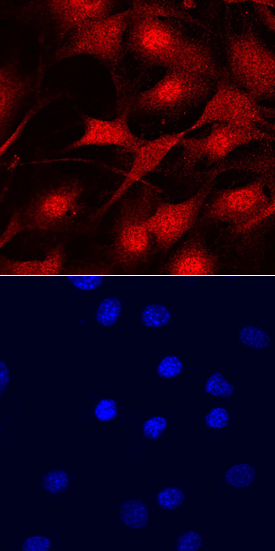
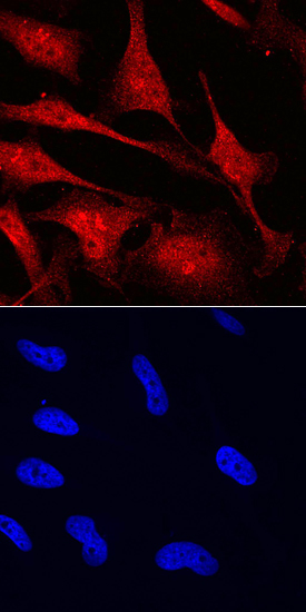
- MYCL1/L-Myc in HeLa Human Cell Line. MYCL1/L-Myc was detected in immersion fixed HeLa human cervical epithelial carcinoma cell line using Goat Anti-Human/Mouse MYCL1/L-Myc Antigen Affinity-purified Polyclonal Antibody (Catalog # AF4050) at 10 µg/mL for 3 hours at room temperature. Cells were stained using the NorthernLights™ 557-conjugated Anti-Goat IgG Secondary Antibody (red, upper panel; Catalog # NL001) and counterstained with DAPI (blue, blue panel). Specific staining was localized to cytoplasm and nuclei. View our protocol for Fluorescent ICC Staining of Cells on Coverslips.
- Submitted by
- R&D Systems (provider)
- Main image

- Experimental details
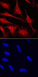
- MYCL1/L-Myc in NIH3T3 Mouse Cell Line. MYCL1/L-Myc was detected in immersion fixed NIH3T3 mouse embryonic fibroblast cell line using Goat Anti-Human/Mouse MYCL1/L-Myc Antigen Affinity-purified Polyclonal Antibody (Catalog # AF4050) at 10 µg/mL for 3 hours at room temperature. Cells were stained using the Northern-Lights™ 557-conjugated Anti-Goat IgG Secondary Antibody (red, upper panel; Catalog # NL001) and counterstained with DAPI (blue, lower panel). Specific staining was localized to cytoplasm and nuclei. View our protocol for Fluorescent ICC Staining of Cells on Coverslips.