Explore
 Validate
 Learn
Learn Western blot
 ELISA
ELISAAntibody data
- Antibody Data
- Antigen structure
- References [0]
- Comments [0]
- Validations
- Western blot [1]
- Immunocytochemistry [1]
Submit
Validation data
Reference
Comment
Report error
- Product number
- R1562P - Provider product page

- Provider
- Acris Antibodies GmbH
- Proper citation
- Acris Antibodies GmbH Cat#R1562P, RRID:AB_1006163
- Product name
- anti CBX4 / PC2
- Antibody type
- Polyclonal
- Antigen
- This affinity purified antibody was prepared from whole rabbit serum produced by repeated immunizations with a synthetic peptide corresponding to aa 95-107 of Human PC2 protein.
- Reactivity
- Human, Mouse
- Host
- Rabbit
- Vial size
- 0.1 mg
- Concentration
- 0.8 mg/ml (by UV absorbance at 280 nm)
No comments: Submit comment
Supportive validation
- Submitted by
- Acris Antibodies GmbH (provider)
- Main image

- Experimental details
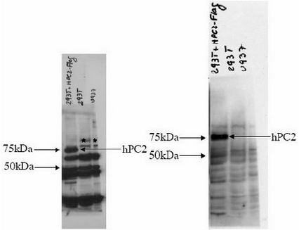
- Figure 2. Western blotting. Analysis shows the detection of human PC2 in probed lysates using Affinity Purified anti-hPC2 antibody. The panel on the left shows the blot probed with anti-hPC2. The panel on the right is the same blot reprobed with anti-FLAG antibody to confirm the presence of FLAG tagged recombinant PC2 in the lysate. In the left panel the band labeled as hPC2 is FLAG-tagged transfected hPC2 in 293T cells. The bands labeled with stars are likely endogenous hPC2. In the right panel the band labeled hPC2 is FLAG-tagged transfected hPC2 in 293T cells. Data contributed by Dr Ari Melnick, Albert Einstein College of Medicine.
Supportive validation
- Submitted by
- Acris Antibodies GmbH (provider)
- Main image
- Experimental details
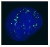
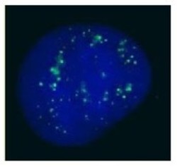
- Figure 1. Immunofluorescence Microscopy. Antibody anti-hPC2 was used for immunofluorescent imaging of human cells (U2OS). The image reveals the expected discrete nuclear structure that is termed the PcG body corresponding to the known localization of PC2 (see Satijn et al. below). IF was performed after fixation in PBS with 4% PF for 5 min, permeabilization with 0.5% Triton X100-PBS for 5 min, and blocking with 5% milk / 0.2% Tween for 1 h. Primary antibody used at 1:200 in 5% milk / 0.2% Tween for 1 h, secondary antibody for 30 min. All blocking and incubation steps carried out at 37°C. Nuclei were counterstained with Hoechst stain (blue). Data contributed by Luke Hughes-Davies and Rhiannon Jade, Gurdon Institute, Cambridge, UK.