Explore
 Validate
 Learn
Learn Western blot
Antibody data
- Antibody Data
- Antigen structure
- References [4]
- Comments [0]
- Validations
- Western blot [1]
- Immunocytochemistry [3]
- Immunohistochemistry [1]
- Flow cytometry [1]
- Chromatin Immunoprecipitation [2]
- Other assay [1]
Submit
Validation data
Reference
Comment
Report error
- Product number
- PA1-802 - Provider product page

- Provider
- Invitrogen Antibodies
- Product name
- FOXL2 Polyclonal Antibody
- Antibody type
- Polyclonal
- Antigen
- Synthetic peptide
- Description
- PA1-802 detects FoxL2 protein in mouse and hamster samples. PA1-802 has successfully been used in Western blot , immunofluorescence and immunohistochemistry procedures. By Western blot, this antibody detects an ~39 kDa protein representing FozL2 in CHO whole cell lysate. The PA1-802 immunogen is a synthetic Peptide corresponding to residues M(1) M A S Y P E P E D T A G T(14) of mouse FoxL2. This sequence is completely conserved in mouse and rat and 92% conserved in human. This peptide (Cat. # PEP-238) is available for use in neutralization and control experiments.
- Reactivity
- Human, Mouse, Hamster
- Host
- Rabbit
- Isotype
- IgG
- Vial size
- 100 μg
- Concentration
- 1 mg/mL
- Storage
- -20°C, Avoid Freeze/Thaw Cycles
Submitted references Etiology of craniofacial malformations in mouse models of blepharophimosis, ptosis and epicanthus inversus syndrome.
Molecular identification and expression of the Foxl2 gene during gonadal sex differentiation in northern snakehead Channa argus.
Molecular cloning and analysis of gonadal expression of Foxl2 in the rice-field eel Monopterus albus.
Foxl2, a forkhead transcription factor, modulates nonclassical activity of the estrogen receptor-alpha.
Heude É, Bellessort B, Fontaine A, Hamazaki M, Treier AC, Treier M, Levi G, Narboux-Nême N
Human molecular genetics 2015 Mar 15;24(6):1670-81
Human molecular genetics 2015 Mar 15;24(6):1670-81
Molecular identification and expression of the Foxl2 gene during gonadal sex differentiation in northern snakehead Channa argus.
Wang DD, Zhang GR, Wei KJ, Ji W, Gardner JP, Yang RB, Chen KC
Fish physiology and biochemistry 2015 Dec;41(6):1419-33
Fish physiology and biochemistry 2015 Dec;41(6):1419-33
Molecular cloning and analysis of gonadal expression of Foxl2 in the rice-field eel Monopterus albus.
Hu Q, Guo W, Gao Y, Tang R, Li D
Scientific reports 2014 Nov 3;4:6884
Scientific reports 2014 Nov 3;4:6884
Foxl2, a forkhead transcription factor, modulates nonclassical activity of the estrogen receptor-alpha.
Kim SY, Weiss J, Tong M, Laronda MM, Lee EJ, Jameson JL
Endocrinology 2009 Nov;150(11):5085-93
Endocrinology 2009 Nov;150(11):5085-93
No comments: Submit comment
Supportive validation
- Submitted by
- Invitrogen Antibodies (provider)
- Main image
- Experimental details
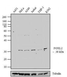
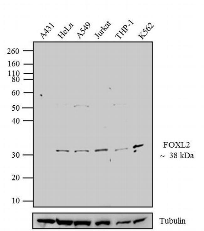
- Western blot analysis was performed on whole cell extracts (30 µg lysate) of A431 (Lane 1), HeLa (Lane 2), A549 (Lane 3), Jurkat (Lane 4), THP-1 (Lane 5) and K562 (Lane 6). The blots were probed with Anti-FOXL2 Rabbit Polyclonal Antibody (Product # PA1-802, 0.1-1 µg/mL) and detected by chemiluminescence using Goat anti-Rabbit IgG (H+L) Secondary Antibody, HRP conjugate (Product # G-21234, 1:5000 dilution). A 38 kDa band corresponding to FOXL2 was observed across cell lines tested except A431. Known quantity of protein samples were electrophoresed using Novex® NuPAGE® 12 % Bis-Tris gel (Product # NP0342BOX), XCell SureLock™ Electrophoresis System (Product # EI0002) and Novex® Sharp Pre-Stained Protein Standard (Product # LC5800). Resolved proteins were then transferred onto a nitrocellulose membrane with iBlot® 2 Dry Blotting System (Product # IB21001). The membrane was probed with the relevant primary and secondary Antibody following blocking with 5 % skimmed milk. Chemiluminescent detection was performed using Pierce™ ECL Western Blotting Substrate (Product # 32106).
Supportive validation
- Submitted by
- Invitrogen Antibodies (provider)
- Main image
- Experimental details
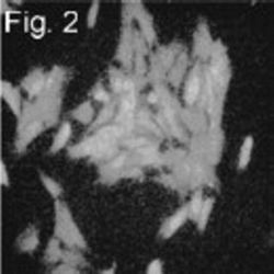
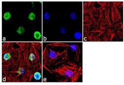
- Immunofluorescence of FoxL2 in KK1 granulosa cells using Product # PA1-802.

- Submitted by
- Invitrogen Antibodies (provider)
- Main image

- Experimental details
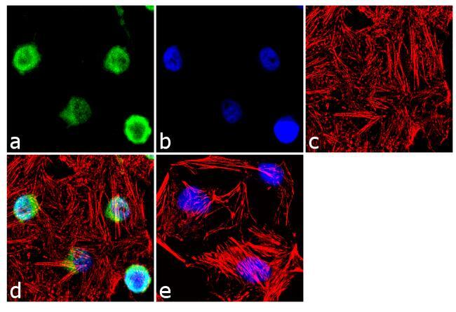
- Immunofluorescence analysis of FOXL2 was done on 70% confluent log phase HeLa cells. The cells were fixed with 4% paraformaldehyde for 10 minutes, permeabilized with 0.1% Triton™ X-100 for 10 minutes, and blocked with 1% BSA for 1 hour at room temperature. The cells were labeled with FOXL2 Rabbit Polyclonal Antibody (Product # PA1-802) at 2 µg/mL in 0.1% BSA and incubated for 3 hours at room temperature and then labeled with Goat anti-Rabbit IgG (H+L) Superclonal™ Secondary Antibody, Alexa Fluor® 488 conjugate (Product # A27034) at a dilution of 1:2000 for 45 minutes at room temperature (Panel a: green). Nuclei (Panel b: blue) were stained with SlowFade® Gold Antifade Mountant with DAPI (Product # S36938). F-actin (Panel c: red) was stained with Alexa Fluor® 555 Rhodamine Phalloidin (Product # R415, 1:300). Panel d is a merged image showing nuclear localization. Panel e is a no primary antibody control. The images were captured at 60X magnification.

- Submitted by
- Invitrogen Antibodies (provider)
- Main image
- Experimental details
- Immunofluorescence analysis of FOXL2 was done on 70% confluent log phase HeLa cells. The cells were fixed with 4% paraformaldehyde for 10 minutes, permeabilized with 0.1% Triton™ X-100 for 10 minutes, and blocked with 1% BSA for 1 hour at room temperature. The cells were labeled with FOXL2 Rabbit Polyclonal Antibody (Product # PA1-802) at 2 µg/mL in 0.1% BSA and incubated for 3 hours at room temperature and then labeled with Goat anti-Rabbit IgG (Heavy Chain) Superclonal™ Secondary Antibody, Alexa Fluor® 488 conjugate (Product # A27034) at a dilution of 1:2000 for 45 minutes at room temperature (Panel a: green). Nuclei (Panel b: blue) were stained with SlowFade® Gold Antifade Mountant with DAPI (Product # S36938). F-actin (Panel c: red) was stained with Alexa Fluor® 555 Rhodamine Phalloidin (Product # R415, 1:300). Panel d is a merged image showing nuclear localization. Panel e is a no primary antibody control. The images were captured at 60X magnification.
Supportive validation
- Submitted by
- Invitrogen Antibodies (provider)
- Main image
- Experimental details
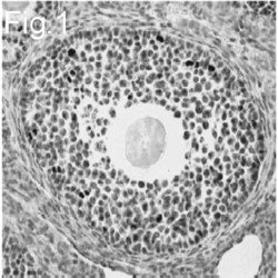
- Immunohistochemical staining of FoxL2 in murine prenatal follicle using Product # PA1-802.

Supportive validation
- Submitted by
- Invitrogen Antibodies (provider)
- Main image
- Experimental details
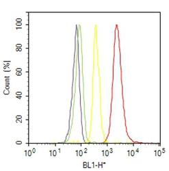
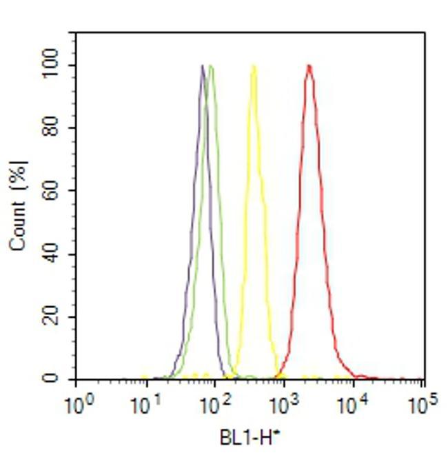
- Flow cytometry analysis of FOXL2 was done on A-431 cells. Cells were fixed with 70% ethanol for 10 minutes, permeabilized with 0.25% Triton™ X-100 for 20 minutes, and blocked with 5% BSA for 30 minutes at room temperature. Cells were labeled with FOXL2 Rabbit Polyclonal Antibody (PA1802, red histogram) or with rabbit isotype control (yellow histogram) at 3-5 ug/million cells in 2.5% BSA. After incubation at room temperature for 2 hours, the cells were labeled with Alexa Fluor® 488 Goat Anti-Rabbit Secondary Antibody (A11008) at a dilution of 1:400 for 30 minutes at room temperature. The representative 10,000 cells were acquired and analyzed for each sample using an Attune® Acoustic Focusing Cytometer. The purple histogram represents unstained control cells and the green histogram represents no-primary-antibody control.
Supportive validation
- Submitted by
- Invitrogen Antibodies (provider)
- Main image
- Experimental details
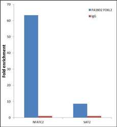
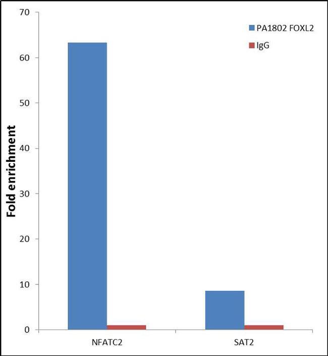
- Enrichment of endogenous FOXL2 Protein at specific gene loci using Anti-FOXL2 Protein Rabbit Polyclonal Antibody: Chromatin Immunoprecipitation (ChIP) was performed using Anti-FOXL2 Protein Rabbit Polyclonal Antibody (Product # PA1-802, 3 µg) on sheared chromatin from 2 million HeLa cells using the "MAGnify ChIP system" kit (Product # 49-2024). Normal Rabbit IgG was used as a negative IP control. The purified DNA was analyzed by 7500 Fast qPCR system (Product # 4351106) with optimized PCR primer pairs for the promoter of active NFATC2 gene, used as positive control target, and the SAT2, used as negative control target. Data is presented as fold enrichment of the antibody signal versus the negative control IgG using the comparative CT method.

- Submitted by
- Invitrogen Antibodies (provider)
- Main image

- Experimental details
- Enrichment of endogenous FOXL2 Protein at specific gene loci using Anti-FOXL2 Protein Rabbit Polyclonal Antibody: Chromatin Immunoprecipitation (ChIP) was performed using Anti-FOXL2 Protein Rabbit Polyclonal Antibody (Product # PA1-802, 3 µg) on sheared chromatin from 2 million HeLa cells using the "MAGnify ChIP system" kit (Product # 49-2024). Normal Rabbit IgG was used as a negative IP control. The purified DNA was analyzed by 7500 Fast qPCR system (Product # 4351106) with optimized PCR primer pairs for the promoter of active NFATC2 gene, used as positive control target, and the SAT2, used as negative control target. Data is presented as fold enrichment of the antibody signal versus the negative control IgG using the comparative CT method.
Supportive validation
- Submitted by
- Invitrogen Antibodies (provider)
- Main image
- Experimental details
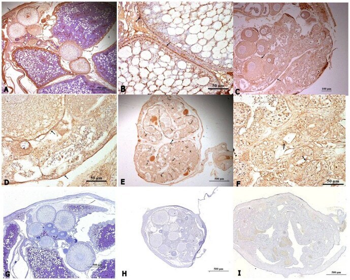
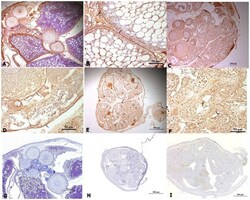
- Figure 7 Immunohistochemical analysis of Foxl2 in different phases of M. albus gonadal development. (A-F) Immunohistochemical analysis of Foxl2 in the (A and B) ovaries, (C and D) ovotestis, and (E and F) testis. (B), (D), and (F) are enlarged areas of (A), (C), and (E), respectively. The positive antigen was dyed brown with 3',3'-diaminobenzidine (DAB) (arrows). (G-I) Negative controls.