Explore
 Validate
 Learn
Learn Western blot
 Immunocytochemistry
Antibody data
- Antibody Data
- Antigen structure
- References [3]
- Comments [0]
- Validations
- Immunocytochemistry [1]
- Chromatin Immunoprecipitation [1]
Submit
Validation data
Reference
Comment
Report error
- Product number
- HPA034488 - Provider product page

- Provider
- Atlas Antibodies
- Proper citation
- Atlas Antibodies Cat#HPA034488, RRID:AB_10669783
- Product name
- Anti-LDB1
- Antibody type
- Polyclonal
- Description
- Polyclonal Antibody against Human LDB1, Gene description: LIM domain binding 1, Alternative Gene Names: CLIM2, NLI, Validated applications: ICC, ChIP, IHC, WB, Uniprot ID: Q86U70, Storage: Store at +4°C for short term storage. Long time storage is recommended at -20°C.
- Reactivity
- Human
- Host
- Rabbit
- Conjugate
- Unconjugated
- Isotype
- IgG
- Vial size
- 100 µl
- Concentration
- 0.4 mg/ml
- Storage
- Store at +4°C for short term storage. Long time storage is recommended at -20°C.
- Handling
- The antibody solution should be gently mixed before use.
Submitted references Ldb1 and Rnf12-dependent regulation of Lhx2 controls the relative balance between neurogenesis and gliogenesis in retina
Immunofluorescence and fluorescent-protein tagging show high correlation for protein localization in mammalian cells
The interactome of LIM domain proteins: The contributions of LIM domain proteins to heart failure and heart development
de Melo J, Clark B, Venkataraman A, Shiau F, Zibetti C, Blackshaw S
Development 2018
Development 2018
Immunofluorescence and fluorescent-protein tagging show high correlation for protein localization in mammalian cells
Stadler C, Rexhepaj E, Singan V, Murphy R, Pepperkok R, Uhlén M, Simpson J, Lundberg E
Nature Methods 2013;10(4):315-323
Nature Methods 2013;10(4):315-323
The interactome of LIM domain proteins: The contributions of LIM domain proteins to heart failure and heart development
Li A, Ponten F, dos Remedios C
PROTEOMICS 2012;12(2):203-225
PROTEOMICS 2012;12(2):203-225
No comments: Submit comment
Supportive validation
- Submitted by
- Atlas Antibodies (provider)
- Main image

- Experimental details
- Immunofluorescent staining of human cell line A-431 shows localization to nucleoplasm.
- Sample type
- Human

Supportive validation
- Submitted by
- Atlas Antibodies (provider)
- Main image

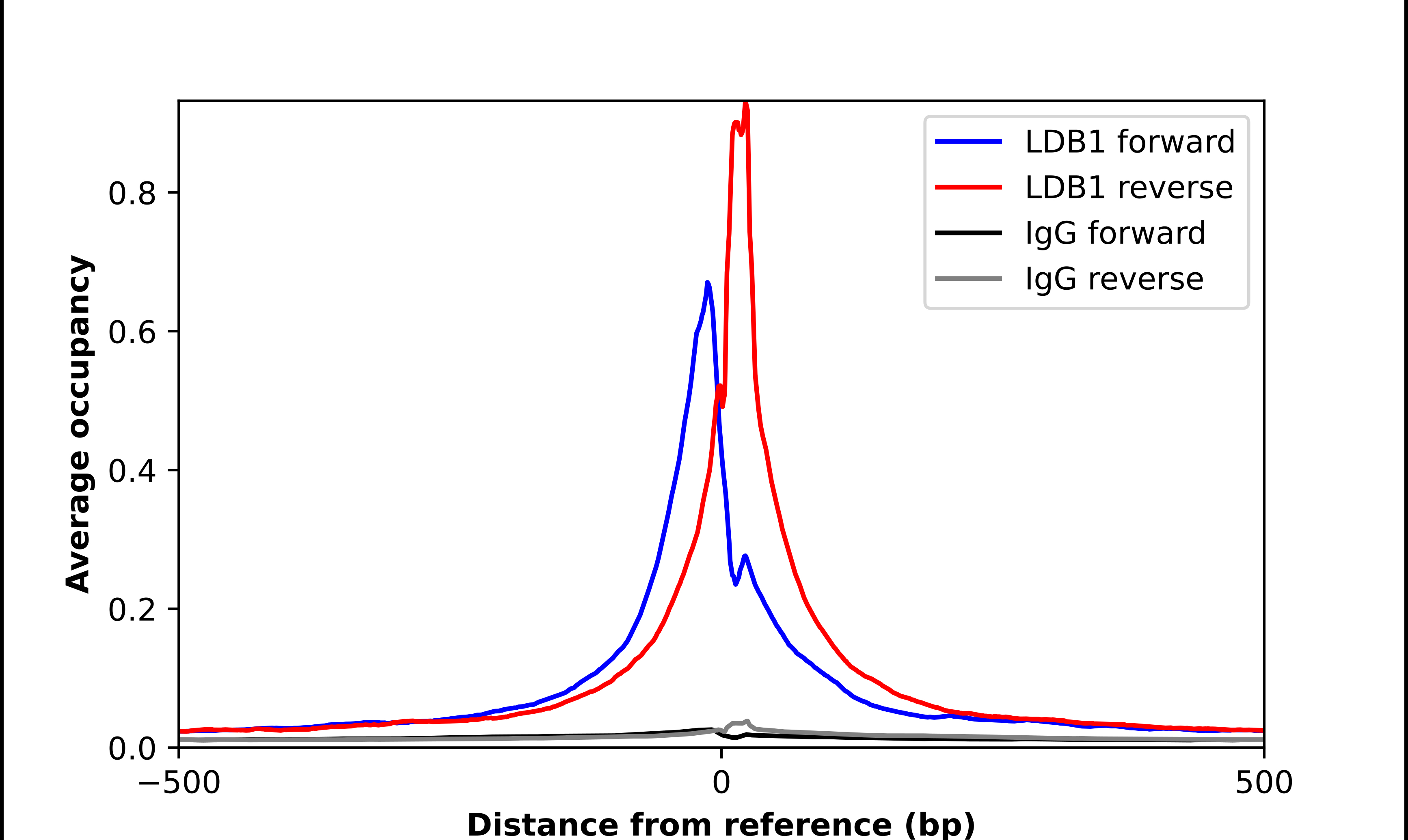
- Experimental details
- ChIP-Exo-Seq composite graph for Anti-LDB1 (HPA034488, Lot A118406) tested in K562 cells. Strand-specific reads (blue: forward, red: reverse) and IgG controls (black: forward, grey: reverse) are plotted against the distance from a composite set of reference binding sites. The antibody exhibits robust target enrichment compared to a non-specific IgG control and precisely reveals its structural organization around the binding site. Data generated by Prof. B. F. Pugh´s Lab at Cornell University.