 Explore
 Validate
 Learn
Learn Western blot
Antibody data
- Antibody Data
- Antigen structure
- References [0]
- Comments [0]
- Validations
- Western blot [3]
- Immunocytochemistry [1]
- Immunohistochemistry [1]
Submit
Validation data
Reference
Comment
Report error
- Product number
- PA5-56948 - Provider product page

- Provider
- Invitrogen Antibodies
- Product name
- LDB1 Polyclonal Antibody
- Antibody type
- Polyclonal
- Antigen
- Recombinant full-length protein
- Description
- Immunogen sequence: MLDRDVGPTP MYPPTYLEPG IGRHTPYGNQ TDYRIFELNK RLQNWTEECD NLW
- Concentration
- 0.50 mg/mL
No comments: Submit comment
Supportive validation
- Submitted by
- Invitrogen Antibodies (provider)
- Main image
- Experimental details
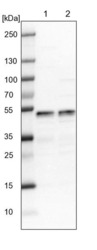
- Western blot analysis of LDB1 in Lane 1: NIH-3T3 cell lysate (Mouse embryonic fibroblast cells); Lane 2: NBT-II cell lysate (Rat Wistar bladder tumour cells). Samples were probed using a LDB1 Polyclonal Antibody (Product # PA5-56948).

- Submitted by
- Invitrogen Antibodies (provider)
- Main image

- Experimental details
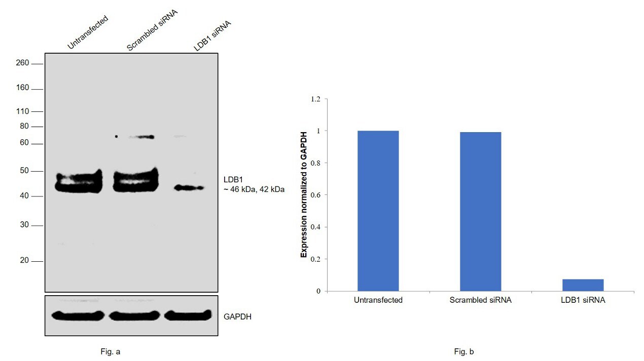
- Knockdown of LDB1 was achieved by transfecting HeLa with LDB1 specific siRNAs (Silencer® select Product # s16921). Western blot analysis (Fig. a) was performed using whole cell extracts from the LDB1 knockdown cells (lane 3), non-specific scrambled siRNA transfected cells (lane 2) and untransfected cells (lane 1). The blot was probed with LDB1 Polyclonal Antibody (Product # PA5-56948, 0.4ug/mL dilution) and Goat anti-Rabbit IgG (H+L) Superclonal™ Secondary Antibody, HRP (Product # A27036, 0.25µg/ml, 1:4000 dilution). Densitometric analysis of this western blot is shown in histogram (Fig. b). Decrease in signal upon siRNA mediated knock down confirms that antibody is specific to LDB1.
- Submitted by
- Invitrogen Antibodies (provider)
- Main image
- Experimental details
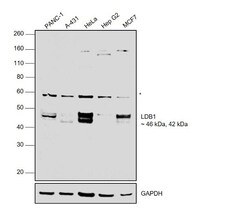
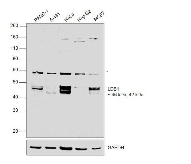
- Western blot was performed using LDB1 Rabbit Polyclonal Antibody (Product # PA5-56948) and 46kDa, 42kDa bands corresponding to LDB1 was observed across cell lines along with an uncharacterized band (*) at ~60kDa in the cell lines tested. Modified whole cell extracts (1% SDS) (30 µg lysate) of PANC-1 (Lane 1), A-431 (Lane 2), HeLa (Lane 3), Hep G2 (Lane 4) and MCF7 (Lane 5) were electrophoresed using NuPAGE® 4-12 % Bis-Tris gel (Product # NP0322BOX). Resolved proteins were then transferred onto a nitrocellulose membrane (Product # IB23001) by iBlot® 2 Dry Blotting System (Product # IB21001).The blot was probed with the primary antibody (0.4ug/mL) and detected by chemiluminescence Goat Anti-Rabbit IgG Secondary Antibody, HRP conjugate (Product # A27036, 1:4000 dilution) using the iBright FL 1000 (Product # A32752). Chemiluminescent detection was performed using Novex® ECL Chemiluminescent Substrate Reagent Kit (Product # WP20005).
Supportive validation
- Submitted by
- Invitrogen Antibodies (provider)
- Main image

- Experimental details
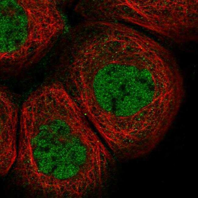
- Immunofluorescent staining of LDB1 in human cell line A-431 shows positivity in nucleus but excluded from the nucleoli. Samples were probed using a LDB1 Polyclonal Antibody (Product # PA5-56948).
Supportive validation
- Submitted by
- Invitrogen Antibodies (provider)
- Main image

- Experimental details
- Immunohistochemical staining of LDB1 in human stomach tissue shows distinct nuclear and cytoplasmic positivity in glandular cells. Samples were probed using a LDB1 Polyclonal Antibody (Product # PA5-56948).
