 Explore
 Validate
 Learn
Learn Western blot
Antibody data
- Antibody Data
- Antigen structure
- References [3]
- Comments [0]
- Validations
- Western blot [1]
Submit
Validation data
Reference
Comment
Report error
- Product number
- PAB10257 - Provider product page

- Provider
- Abnova Corporation
- Proper citation
- Abnova Corporation Cat#PAB10257, RRID:AB_1676512
- Product name
- Ldb1 polyclonal antibody
- Antibody type
- Polyclonal
- Description
- Rabbit polyclonal antibody raised against synthetic peptide of Ldb1.
- Storage
- Store at 4°C. For long term storage store at -20°C.Aliquot to avoid repeated freezing and thawing.
Submitted references Identification of a TAL1 target gene reveals a positive role for the LIM domain-binding protein Ldb1 in erythroid gene expression and differentiation.
Functional ablation of the mouse Ldb1 gene results in severe patterning defects during gastrulation.
Genomic structure and chromosomal localization of the mouse LIM domain-binding protein 1 gene, Ldb1.
Xu Z, Huang S, Chang LS, Agulnick AD, Brandt SJ
Molecular and cellular biology 2003 Nov;23(21):7585-99
Molecular and cellular biology 2003 Nov;23(21):7585-99
Functional ablation of the mouse Ldb1 gene results in severe patterning defects during gastrulation.
Mukhopadhyay M, Teufel A, Yamashita T, Agulnick AD, Chen L, Downs KM, Schindler A, Grinberg A, Huang SP, Dorward D, Westphal H
Development (Cambridge, England) 2003 Feb;130(3):495-505
Development (Cambridge, England) 2003 Feb;130(3):495-505
Genomic structure and chromosomal localization of the mouse LIM domain-binding protein 1 gene, Ldb1.
Yamashita T, Agulnick AD, Copeland NG, Gilbert DJ, Jenkins NA, Westphal H
Genomics 1998 Feb 15;48(1):87-92
Genomics 1998 Feb 15;48(1):87-92
No comments: Submit comment
Supportive validation
- Submitted by
- Abnova Corporation (provider)
- Main image
- Experimental details
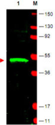
- Western blot using Ldb1 polyclonal antibody (Cat # PAB10257) shows detection of Ldb1 protein (arrowhead) in Jurkat whole cell lysate.Approximately 30 ug of lysate was loaded prior to separation and transfer to nitrocellulose.Primary antibody was used at a 1 : 1,800 dilution in 5% BLOTTO in PBS reacted overnight at 4°C.The membrane was washed and reacted with a 1:20,000 dilution of DyLight™800 conjugated Gt-a-Rabbit IgG [H&L] MX for 45 min at room temperature (800 nm channel, green).Molecular weight estimation was made by comparison to prestained MW markers in lane M (700 nm channel, red).Fluorescence image was captured using the Odyssey® Infrared Imaging System developed by LI-COR.IRDye is a trademark of LI-COR, Inc.
