 Explore
 Validate
 Learn
Learn Western blot
 Immunocytochemistry
Antibody data
- Antibody Data
- Antigen structure
- References [1]
- Comments [0]
- Validations
- Western blot [3]
- Immunohistochemistry [1]
Submit
Validation data
Reference
Comment
Report error
- Product number
- NBP2-20132 - Provider product page

- Provider
- Novus Biologicals
- Product name
- Rabbit Polyclonal Retinol Binding Protein RBP Antibody
- Antibody type
- Polyclonal
- Description
- Immunogen affinity purified.
- Reactivity
- Human, Mouse
- Host
- Rabbit
- Isotype
- IgG
- Vial size
- 0.1 ml
- Storage
- Aliquot and store at -20C or -80C. Avoid freeze-thaw cycles.
Submitted references Chrysin Ameliorates Malfunction of Retinoid Visual Cycle through Blocking Activation of AGE-RAGE-ER Stress in Glucose-Stimulated Retinal Pigment Epithelial Cells and Diabetic Eyes.
Kang MK, Lee EJ, Kim YH, Kim DY, Oh H, Kim SI, Kang YH
Nutrients 2018 Aug 8;10(8)
Nutrients 2018 Aug 8;10(8)
No comments: Submit comment
Supportive validation
- Submitted by
- Novus Biologicals (provider)
- Main image

- Experimental details
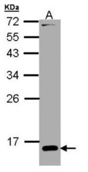
- Western Blot: Retinol Binding Protein RBP Antibody [NBP2-20132] - CRBP I antibody detects RBP1 protein by Western blot analysis. A. 30 ug NIH-3T3 whole cell lysate/extractB. 30 ug C2C12 whole cell lysate/extract12 % SDS-PAGECRBP I antibody dilution: 1:5000.

- Submitted by
- Novus Biologicals (provider)
- Main image
- Experimental details
- Western Blot: Retinol Binding Protein RBP Antibody [NBP2-20132] - Sample (30 ug of whole cell lysate) A: H1299 15% SDS PAGE gel, diluted at 1:5000.

- Submitted by
- Novus Biologicals (provider)
- Main image

- Experimental details
- Western Blot: Retinol Binding Protein RBP Antibody [NBP2-20132] - Elevation of retinal tissue induction of visual cycle enzymes stimulated by retinoic acid 6 (STRA6, C) in chrysin-treated mice. The db/db mice were orally supplemented with 10 mg/kg of chrysin daily for 10 weeks. The db/m mice were introduced as control animals. Mouse retinal tissue extracts were subject to Western blot analysis with a primary antibody against each target protein of LRAT, RDH5, cellular retinol binding protein (CRBP), CRALBP, IRBP, or STRA6. beta-actin protein was used as an internal control. Bar graphs (mean +- SEM, n = nine independent experiments) in the bottom or right panels represent densitometric results of upper blot bands. * Values in bar graphs indicate a significant difference at p

Supportive validation
- Submitted by
- Novus Biologicals (provider)
- Main image
- Experimental details
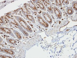
- Immunohistochemistry-Paraffin: Retinol Binding Protein RBP Antibody [NBP2-20132] - Human colon, at 1:100 dilution. Antigen Retrieval: Trilogy™ (EDTA based, pH 8.0) buffer, 15min
