Explore
 Validate
 Learn
Learn Immunohistochemistry
Antibody data
- Antibody Data
- Antigen structure
- References [0]
- Comments [0]
- Validations
- Immunohistochemistry [1]
- Flow cytometry [1]
- Blocking/Neutralizing [1]
Submit
Validation data
Reference
Comment
Report error
- Product number
- MAB8210 - Provider product page

- Provider
- Novus Biologicals
- Product name
- Mouse Monoclonal GIPR Antibody
- Antibody type
- Monoclonal
- Description
- Protein A or G purified from hybridoma culture supernatant. Detects human GIPR in direct ELISAs. Stains human GIPR transfected cells but not irrelevant transfectants.
- Reactivity
- Human
- Host
- Mouse
- Conjugate
- Unconjugated
- Isotype
- IgG
- Vial size
- 100 ug
- Concentration
- LYOPH
- Storage
- Use a manual defrost freezer and avoid repeated freeze-thaw cycles. 12 months from date of receipt, -20 to -70 degreesC as supplied. 1 month, 2 to 8 degreesC under sterile conditions after reconstitution. 6 months, -20 to -70 degreesC under sterile conditions after reconstitution.
No comments: Submit comment
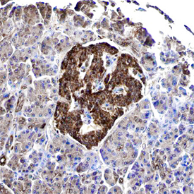
Supportive validation
- Submitted by
- Novus Biologicals (provider)
- Main image
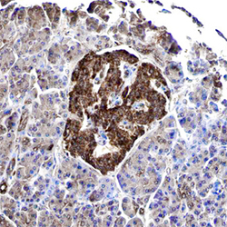
- Experimental details
- GIPR in Human Pancreas. GIPR was detected in immersion fixed paraffin-embedded sections of human pancreas using Mouse Anti-Human GIPR Monoclonal Antibody (Catalog # MAB8210) at 15 µg/mL overnight at 4 °C. Before incubation with the primary antibody, tissue was subjected to heat-induced epitope retrieval using Antigen Retrieval Reagent-Basic (Catalog # CTS013). Tissue was stained using the Anti-Mouse HRP-DAB Cell & Tissue Staining Kit (brown; Catalog # CTS002) and counterstained with hematoxylin (blue). Specific staining was localized to islet cells. View our protocol for Chromogenic IHC Staining of Paraffin-embedded Tissue Sections.
Supportive validation
- Submitted by
- Novus Biologicals (provider)
- Main image
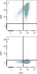
- Experimental details
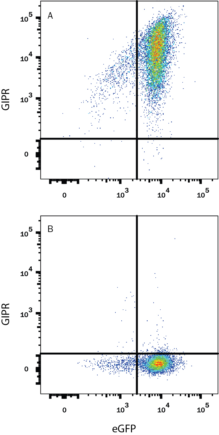
- Detection of GIPR in HEK293 Human Cell Line Transfected with Human GIPR and eGFP by Flow Cytometry. HEK293 human embryonic kidney cell line transfected with either (A) human GIPR or (B) irrelevant transfectants and eGFP were stained with Mouse Anti-Human GIPR Monoclonal Antibody (Catalog # MAB8210) followed by Allophycocyanin-conjugated Anti-Mouse IgG Secondary Antibody (Catalog # F0101B). Quadrant markers were set based on control antibody staining (Catalog # MAB002). View our protocol for Staining Membrane-associated Proteins.
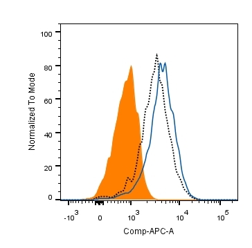
Supportive validation
- Submitted by
- Novus Biologicals (provider)
- Main image

- Experimental details
- Human GIP binding to human GIPR-transfected HEK293 cells blocked by Human GIPR Antibody. In a functional flow cytometry test, biotinylated recombinant Human GIP (Catalog # 2257, 25 ng/mL) binds to Human GIPR-transfected HEK293 cells (black dotted line). Binding is completely blocked (orange histogram) by 2.5 μg/mL of Mouse Anti-Human GIPR Monoclonal Antibody (Catalog # MAB8210). Mouse IgG1 Isotype Control (Catalog # MAB002) at 2.5 μg/mL was used as a control (blue line). Cells were stained with Sterptavidin-APC (Catalog # F0050).