Explore
 Validate
 Learn
Learn Western blot
Antibody data
- Antibody Data
- Antigen structure
- References [1]
- Comments [0]
- Validations
- Western blot [3]
- Immunohistochemistry [1]
- Other assay [1]
Submit
Validation data
Reference
Comment
Report error
- Product number
- PA1-41581 - Provider product page

- Provider
- Invitrogen Antibodies
- Product name
- MTA2 Polyclonal Antibody
- Antibody type
- Polyclonal
- Antigen
- Synthetic peptide
- Description
- This antibody is predicted to react with mouse, rat, canine, bovine, and xenopus samples based on sequence homology.
- Concentration
- 0.5 mg/mL
Submitted references The chromatin remodeling factor CHD5 is a transcriptional repressor of WEE1.
Quan J, Adelmant G, Marto JA, Look AT, Yusufzai T
PloS one 2014;9(9):e108066
PloS one 2014;9(9):e108066
No comments: Submit comment
Supportive validation
- Submitted by
- Invitrogen Antibodies (provider)
- Main image
- Experimental details
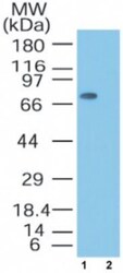
- Western blot analysis of MTA2 in lysate from Jurkat cells, in the 1) absence and 2) presence of immunizing peptide. Samples were incubated in MTA2 polyclonal antibody (Product # PA1-41581) using a dilution of 0.5 µg/mL followed by a goat anti-rabbit Ig HRP secondary antibody. PicoTect ECL substrate solution was used for this test.
- Submitted by
- Invitrogen Antibodies (provider)
- Main image

- Experimental details
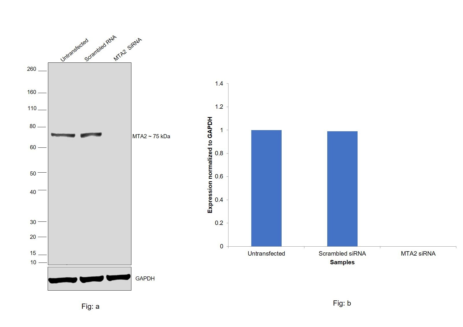
- Knockdown of MTA2 was achieved by transfecting HeLa with MTA2 specific siRNAs (Silencer® select Products # s17390, s17389). Western blot analysis (Fig. a) was performed using whole cell Lysates from the MTA2 knockdown (siRNA treated) cells (Lane 3), non-specific scrambled siRNA transfected cells (Lane 2) and untransfected cells (Lane 1). The blot was probed with MTA2 Polyclonal Antibody (Product # PA1-41581, 1:1000 dilution) and Goat anti-Rabbit IgG (H+L) Superclonal™ Recombinant Secondary Antibody, HRP (Product # A27036, 1:4000 dilution). Densitometric analysis of this western blot is shown in histogram (Fig. b). Decrease in signal upon siRNA mediated knock down confirms that antibody is specific to MTA2.
- Submitted by
- Invitrogen Antibodies (provider)
- Main image
- Experimental details
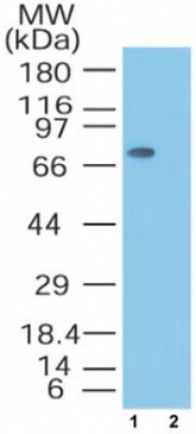
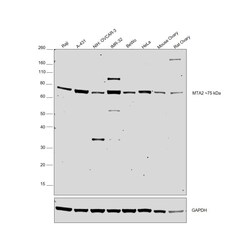
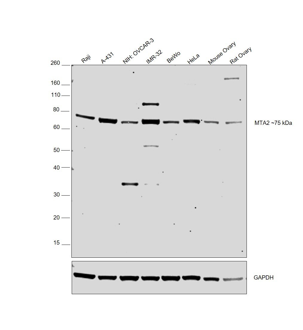
- Western blot was performed using Anti-MTA2 Polyclonal Antibody (Product # PA1-41581) and ~75 kDa band corresponding to MTA2 was observed in all the cell lines and tissues tested. Whole cell lysate (40ug lysate) of Raji (Lane 1), A-431 (Lane 2), NIH: OVCAR-3 (Lane 3), IMR-32 (Lane 4), BeWo (Lane 5), HeLa (Lane 6), Mouse Ovary (Lane 7) and Rat Ovary (Lane 8) were electrophoresed using Novex® NuPAGE® 4-12 % Bis-Tris gel (Product # NP0322BOX). Resolved proteins were then transferred onto a nitrocellulose membrane (Product # IB23001) by iBlot® 2 Dry Blotting System (Product # IB21001). The bot was probed with the primary antibody (1:1000 dilution) and detected by chemiluminescence with Goat anti-Rabbit IgG (H+L) Superclonal™ Recombinant Secondary Antibody, HRP (Product # A27036, 1:4000 dilution) using the iBright FL 1000 (Product # A32752). Chemiluminescent detection was performed using Novex® ECL Chemiluminescent Substrate Reagent Kit (Product # WP20005).
Supportive validation
- Submitted by
- Invitrogen Antibodies (provider)
- Main image
- Experimental details
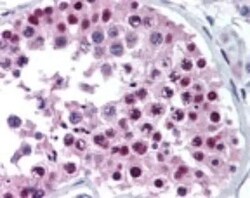
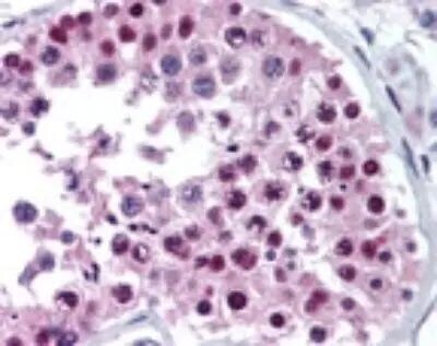
- Immunohistochemical analysis of MTA2 in Human Testis. Samples were incubated in MTA2 polyclonal antibody (Product # PA1-41581) using a dilution of 10 µg/mL.
Supportive validation
- Submitted by
- Invitrogen Antibodies (provider)
- Main image

- Experimental details
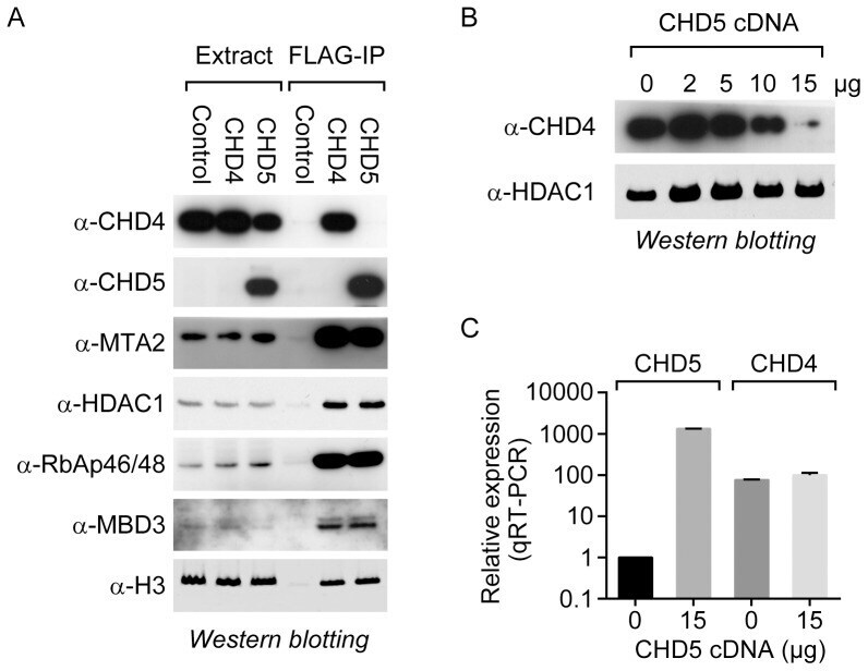
- Figure 1 CHD5 co-purifies with the NuRD transcriptional repressor complex. (A) FLAG-tagged CHD4 and CHD5 were immunoprecipitated from a transiently transfected HEK293T-derived cell line, and the samples were analyzed by western blotting using the indicated antibodies. The empty vector was also transfected in parallel [Control]. (B) The HEK293T-derived line was transiently transfected with increasing amounts of the CHD5-expression plasmid. An empty vector was included where necessary to keep the final amount (15 ug) of transfected plasmid constant. Following transfection, cell extracts and RNA were prepared to measure CHD4 levels by western blotting and CHD4 and CHD5 mRNA levels by qRT-PCR.