 Explore
 Validate
 Learn
Learn Western blot
 ELISA
ELISAAntibody data
- Antibody Data
- Antigen structure
- References [7]
- Comments [0]
- Validations
- Western blot [1]
- Immunocytochemistry [1]
- Immunohistochemistry [2]
Submit
Validation data
Reference
Comment
Report error
- Product number
- 14805-1-AP - Provider product page

- Provider
- Proteintech Group
- Proper citation
- Proteintech Cat#14805-1-AP, RRID:AB_2101837
- Product name
- EXOSC2 antibody
- Antibody type
- Polyclonal
- Description
- KD/KO validated EXOSC2 antibody (Cat. #14805-1-AP) is a rabbit polyclonal antibody that shows reactivity with human and has been validated for the following applications: IF, IHC, IP, WB,ELISA.
- Reactivity
- Human
- Host
- Rabbit
- Conjugate
- Unconjugated
- Isotype
- IgG
- Vial size
- 20ul, 150ul
Submitted references Humanized Saccharomyces cerevisiae provides a facile and effective tool to identify damaging human variants that cause exosomopathies.
EXOSC2 Mediates the Pro-tumor Role of WTAP in Breast Cancer Cells via Activating the Wnt/β-Catenin Signal.
Dual modes of ZFC3H1 confer selectivity in nuclear RNA sorting.
The MYCN oncoprotein is an RNA-binding accessory factor of the nuclear exosome targeting complex.
Low expression of EXOSC2 protects against clinical COVID-19 and impedes SARS-CoV-2 replication.
The Human RNA-Binding Proteome and Its Dynamics during Translational Arrest.
Modulated Expression of Specific tRNAs Drives Gene Expression and Cancer Progression.
Ahammed KS, Fasken MB, Corbett AH, van Hoof A
G3 (Bethesda, Md.) 2025 Apr 17;15(4)
G3 (Bethesda, Md.) 2025 Apr 17;15(4)
EXOSC2 Mediates the Pro-tumor Role of WTAP in Breast Cancer Cells via Activating the Wnt/β-Catenin Signal.
Lv CG, Cheng Y, Zhang L, Wu GG, Liang CY, Tao Z, Chen B
Molecular biotechnology 2024 Sep;66(9):2569-2582
Molecular biotechnology 2024 Sep;66(9):2569-2582
Dual modes of ZFC3H1 confer selectivity in nuclear RNA sorting.
Fan J, Wang Y, Wen M, Tong D, Wu K, Yan K, Jia P, Zhu Y, Liu Q, Zou H, Zhao P, Lu F, Yun C, Xue Y, Zhou Y, Cheng H
Molecular cell 2024 Nov 21;84(22):4297-4313.e7
Molecular cell 2024 Nov 21;84(22):4297-4313.e7
The MYCN oncoprotein is an RNA-binding accessory factor of the nuclear exosome targeting complex.
Papadopoulos D, Ha SA, Fleischhauer D, Uhl L, Russell TJ, Mikicic I, Schneider K, Brem A, Valanju OR, Cossa G, Gallant P, Schuelein-Voelk C, Maric HM, Beli P, Büchel G, Vos SM, Eilers M
Molecular cell 2024 Jun 6;84(11):2070-2086.e20
Molecular cell 2024 Jun 6;84(11):2070-2086.e20
Low expression of EXOSC2 protects against clinical COVID-19 and impedes SARS-CoV-2 replication.
Moll T, Odon V, Harvey C, Collins MO, Peden A, Franklin J, Graves E, Marshall JN, Dos Santos Souza C, Zhang S, Castelli L, Hautbergue G, Azzouz M, Gordon D, Krogan N, Ferraiuolo L, Snyder MP, Shaw PJ, Rehwinkel J, Cooper-Knock J
Life science alliance 2023 Jan;6(1)
Life science alliance 2023 Jan;6(1)
The Human RNA-Binding Proteome and Its Dynamics during Translational Arrest.
Trendel J, Schwarzl T, Horos R, Prakash A, Bateman A, Hentze MW, Krijgsveld J
Cell 2019 Jan 10;176(1-2):391-403.e19
Cell 2019 Jan 10;176(1-2):391-403.e19
Modulated Expression of Specific tRNAs Drives Gene Expression and Cancer Progression.
Goodarzi H, Nguyen HCB, Zhang S, Dill BD, Molina H, Tavazoie SF
Cell 2016 Jun 2;165(6):1416-1427
Cell 2016 Jun 2;165(6):1416-1427
No comments: Submit comment
Supportive validation
- Submitted by
- Proteintech Group (provider)
- Main image
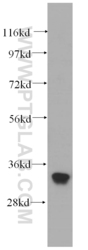
- Experimental details
- HeLa cells were subjected to SDS PAGE followed by western blot with 14805-1-AP(EXOSC2 antibody) at dilution of 1:500
- Sample type
- cell line

Supportive validation
- Submitted by
- Proteintech Group (provider)
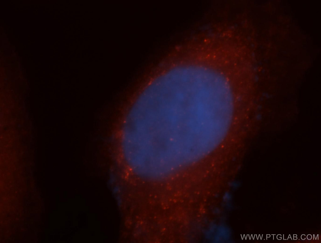
- Main image

- Experimental details
- Immunofluorescent analysis of MCF-7 cells, using EXOSC2 antibody 14805-1-AP at 1:50 dilution and Rhodamine-labeled goat anti-rabbit IgG (red). Blue pseudocolor = DAPI (fluorescent DNA dye).
- Sample type
- cell line
Supportive validation
- Submitted by
- Proteintech Group (provider)
- Main image
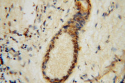
- Experimental details
- Immunohistochemical of paraffin-embedded human skin cancer using 14805-1-AP(EXOSC2 antibody) at dilution of 1:100 (under 40x lens)
- Sample type
- tissue
- Submitted by
- Proteintech Group (provider)
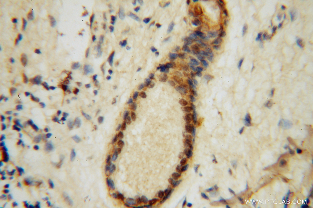
- Main image
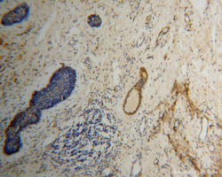
- Experimental details
- Immunohistochemical of paraffin-embedded human skin cancer using 14805-1-AP(EXOSC2 antibody) at dilution of 1:100 (under 10x lens)
- Sample type
- tissue
