 Explore
 Validate
 Learn
Learn Western blot
 Immunocytochemistry
Antibody data
- Antibody Data
- Antigen structure
- References [2]
- Comments [0]
- Validations
- Immunocytochemistry [1]
- Immunohistochemistry [1]
Submit
Validation data
Reference
Comment
Report error
- Product number
- AF908 - Provider product page

- Provider
- R&D Systems
- Product name
- Human MMP-8 Antibody
- Antibody type
- Polyclonal
- Description
- Antigen Affinity-purified. Detects human MMP-8 in direct ELISAs and Western blots. In direct ELISAs, approximately 20% cross-reactivity with recombinant mouse (rm) MMP-8 and recombinant rat MMP-8 is observed, and less than 1% cross-reactivity with recombinant human (rh) MMP-1, rhMMP-2, rhMMP-3, rhMMP-7, rhMMP-9, rhMMP-10, rhMMP-12, rhMMP-13, rmMMP-3, and rmMMP-9 is observed.
- Reactivity
- Human
- Host
- Goat
- Conjugate
- Unconjugated
- Antigen sequence
AAZ38714- Isotype
- IgG
- Vial size
- 100 ug
- Concentration
- LYOPH
- Storage
- Use a manual defrost freezer and avoid repeated freeze-thaw cycles. 12 months from date of receipt, -20 to -70 °C as supplied. 1 month, 2 to 8 °C under sterile conditions after reconstitution. 6 months, -20 to -70 °C under sterile conditions after reconstitution.
Submitted references Redundancy of IL-1 Isoform Signaling and Its Implications for Arterial Remodeling.
Matrix-metalloproteinase-14 deficiency in bone-marrow-derived cells promotes collagen accumulation in mouse atherosclerotic plaques.
Beltrami-Moreira M, Vromman A, Sukhova GK, Folco EJ, Libby P
PloS one 2016;11(3):e0152474
PloS one 2016;11(3):e0152474
Matrix-metalloproteinase-14 deficiency in bone-marrow-derived cells promotes collagen accumulation in mouse atherosclerotic plaques.
Schneider F, Sukhova GK, Aikawa M, Canner J, Gerdes N, Tang SM, Shi GP, Apte SS, Libby P
Circulation 2008 Feb 19;117(7):931-9
Circulation 2008 Feb 19;117(7):931-9
No comments: Submit comment
Supportive validation
- Submitted by
- R&D Systems (provider)
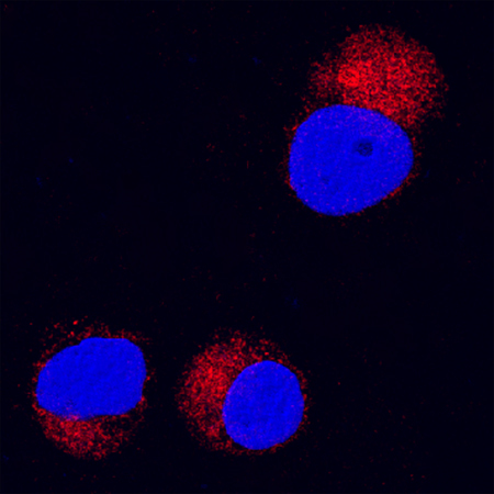
- Main image
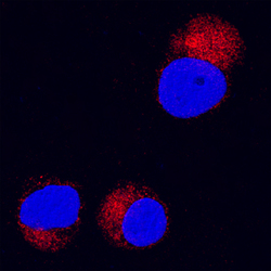
- Experimental details
- MMP-8 in Jurkat Human Cell Line. MMP-8 was detected in immersion fixed Jurkat human acute T cell leukemia cell line using Goat Anti-Human MMP-8 Antigen Affinity-purified Polyclonal Antibody (Catalog # AF908) at 15 µg/mL for 3 hours at room temperature. Cells were stained using the NorthernLights™ 557-conjugated Anti-Goat IgG Secondary Antibody (red; Catalog # NL001) and counterstained with DAPI(blue). Specific staining was localized to cytoplasm. View our protocol for Fluorescent ICC Staining of Cells on Coverslips.
Supportive validation
- Submitted by
- R&D Systems (provider)
- Main image
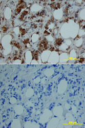
- Experimental details
- MMP-8 in Human Breast. MMP-8 was detected in immersion fixed paraffin-embedded sections of human breast array using Goat Anti-Human MMP-8 Antigen Affinity-purified Polyclonal Antibody (Catalog # AF908) at 15 µg/mL overnight at 4 °C. Tissue was stained using the Anti-Goat HRP-DAB Cell & Tissue Staining Kit (brown; Catalog # CTS008) and counterstained with hematoxylin (blue). Lower panel shows a lack of labeling if primary antibodies are omitted and tissue is stained only with secondary antibody followed by incubation with detection reagents. View our protocol for Chromogenic IHC Staining of Paraffin-embedded Tissue Sections.
