Explore
 Validate
 Learn
Learn Western blot
 Immunoprecipitation
Antibody data
- Antibody Data
- Antigen structure
- References [1]
- Comments [0]
- Validations
- Western blot [1]
- Immunocytochemistry [3]
Submit
Validation data
Reference
Comment
Report error
- Product number
- 73-082 - Provider product page

- Provider
- Antibodies Incorporated / NeuroMab
- Product name
- Anti-Kv7.4/KCNQ4 K+ channel
- Antibody type
- Monoclonal
- Description
- TC Supernatant
- Reactivity
- Human, Mouse, Rat
- Host
- Mouse
- Conjugate
- Unconjugated
- Isotype
- IgG
- Antibody clone number
- N43/6
- Vial size
- 5 mL
- Concentration
- Lot dependent
- Storage
- Aliquot and store at -20 degrees Celsius
Submitted references KCNQ and KCNE potassium channel subunit expression in bovine retinal pigment epithelium.
Zhang X, Hughes BA
Experimental eye research 2013 Nov;116:424-32
Experimental eye research 2013 Nov;116:424-32
No comments: Submit comment
Supportive validation
- Submitted by
- Antibodies Incorporated / NeuroMab (provider)
- Main image
- Experimental details
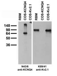
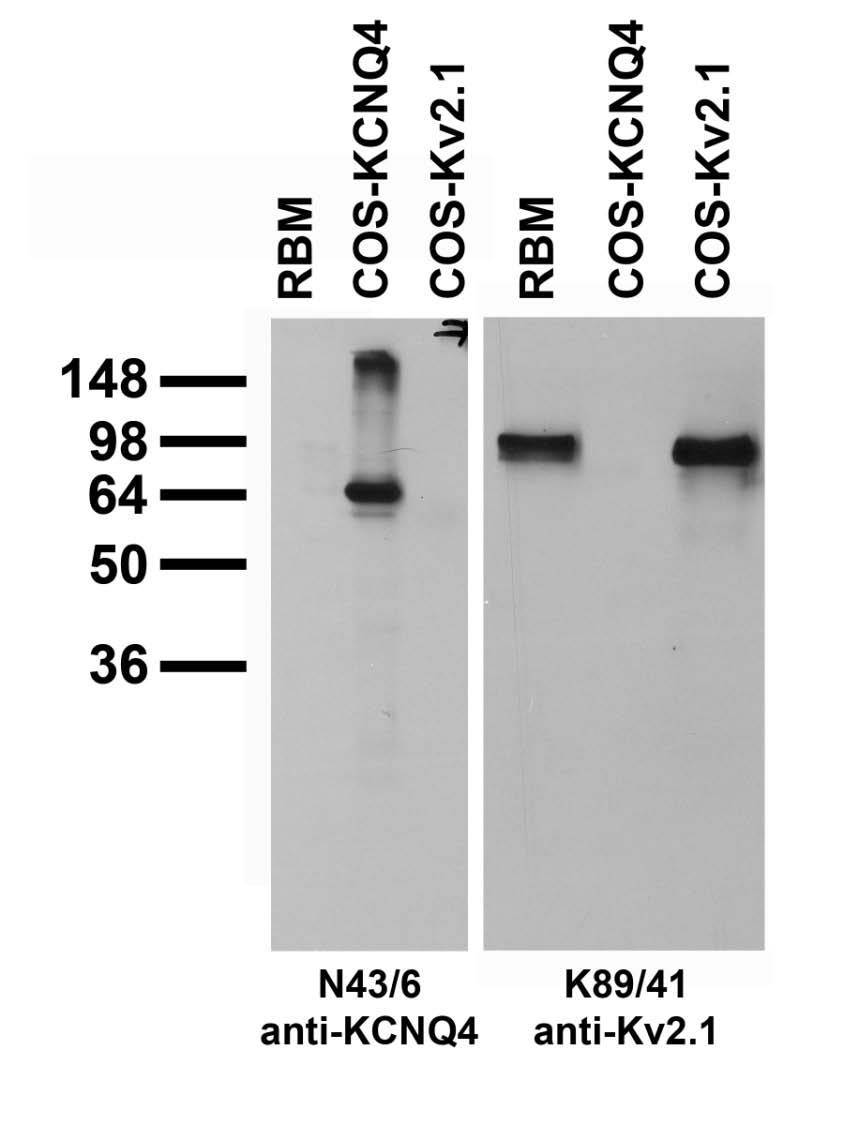
- Adult rat brain membrane (RBM) and transfected cell immunoblot: extracts of RBM (left lane) and COS-1 cells transiently transfected with KCNQ4 (middle lane) or Kv2.1 (right lane) plasmids and probed with pure N43/6 (left panel) or K89/41 TC supe (right panel).
Supportive validation
- Submitted by
- Antibodies Incorporated / NeuroMab (provider)
- Main image

- Experimental details
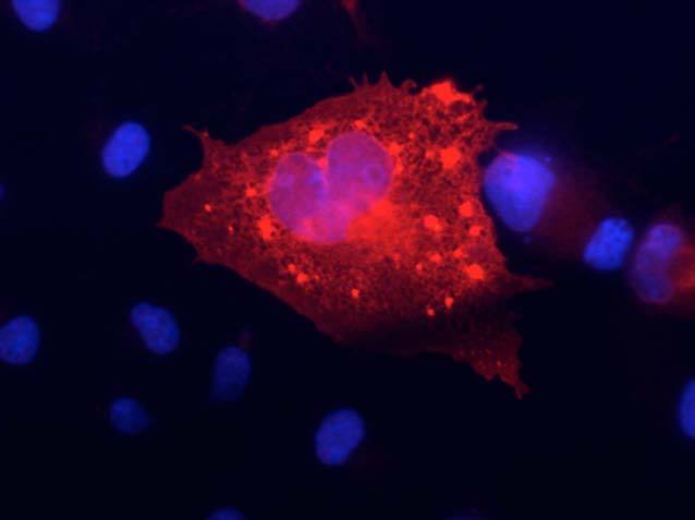
- Transfected cell immunofluorescence :COS-1 cell expressing KCNQ4 in a field of untransfected cells. Red = N43/6, Blue = Hoechst nuclear stain.
- Submitted by
- Antibodies Incorporated / NeuroMab (provider)
- Main image
- Experimental details
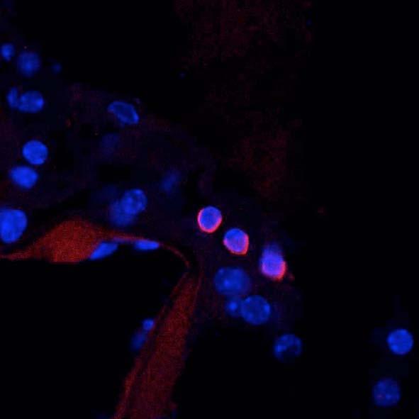
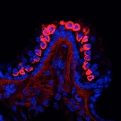
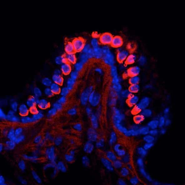
- Mouse cochlea immunofluorescence. Left panel is labeling in crista ampullaris,.
- Submitted by
- Antibodies Incorporated / NeuroMab (provider)
- Main image

- Experimental details
- Right panel in outer hair cells in organ of Corti. Red = N43/6, Blue = Hoechst nuclear stain. Courtesy of Ms. Gesa Rickheit and Dr. Thomas Jentsch, Max-Delbrueck-Centrum fuer Molekulare Medizin, Berlin, Germany.