Explore
 Validate
 Learn
Learn Immunocytochemistry
 Immunohistochemistry
Antibody data
- Antibody Data
- Antigen structure
- References [2]
- Comments [0]
- Validations
- Immunocytochemistry [1]
Submit
Validation data
Reference
Comment
Report error
- Product number
- HPA006092 - Provider product page

- Provider
- Atlas Antibodies
- Proper citation
- Atlas Antibodies Cat#HPA006092, RRID:AB_1079236
- Product name
- Anti-CERS3
- Antibody type
- Polyclonal
- Description
- Polyclonal Antibody against Human CERS3, Gene description: ceramide synthase 3, Alternative Gene Names: LASS3, MGC27091, Validated applications: IHC, ICC, Uniprot ID: Q8IU89, Storage: Store at +4°C for short term storage. Long time storage is recommended at -20°C.
- Reactivity
- Human
- Host
- Rabbit
- Conjugate
- Unconjugated
- Isotype
- IgG
- Vial size
- 100 µl
- Concentration
- 0.4 mg/ml
- Storage
- Store at +4°C for short term storage. Long time storage is recommended at -20°C.
- Handling
- The antibody solution should be gently mixed before use.
Submitted references Mutations in CERS3 Cause Autosomal Recessive Congenital Ichthyosis in Humans
Increased Presence of Monounsaturated Fatty Acids in the Stratum Corneum of Human Skin Equivalents
Wilkie A, Radner F, Marrakchi S, Kirchmeier P, Kim G, Ribierre F, Kamoun B, Abid L, Leipoldt M, Turki H, Schempp W, Heilig R, Lathrop M, Fischer J
PLoS Genetics 2013;9(6):e1003536
PLoS Genetics 2013;9(6):e1003536
Increased Presence of Monounsaturated Fatty Acids in the Stratum Corneum of Human Skin Equivalents
Thakoersing V, van Smeden J, Mulder A, Vreeken R, El Ghalbzouri A, Bouwstra J
Journal of Investigative Dermatology 2013;133(1):59-67
Journal of Investigative Dermatology 2013;133(1):59-67
No comments: Submit comment
Supportive validation
- Submitted by
- Atlas Antibodies (provider)
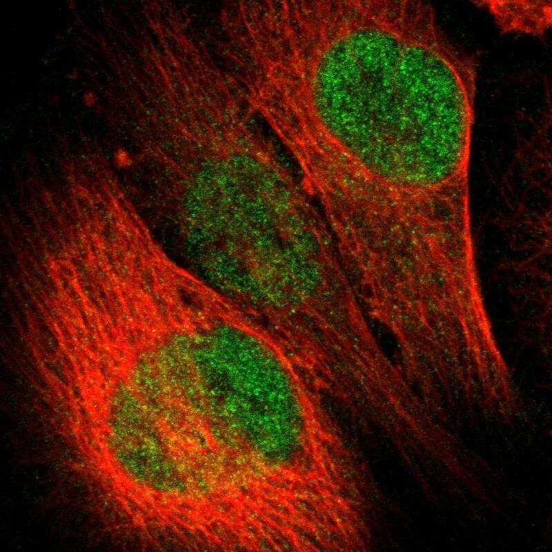
- Main image

- Experimental details
- Immunofluorescent staining of human cell line U-251 MG shows localization to nucleoplasm.
- Sample type
- Human