 Explore
 Validate
 Learn
Learn Western blot
 ELISA
ELISAAntibody data
- Antibody Data
- Antigen structure
- References [0]
- Comments [0]
- Validations
- Western blot [1]
- Immunohistochemistry [1]
Submit
Validation data
Reference
Comment
Report error
- Product number
- AP09286PU-N - Provider product page

- Provider
- Acris Antibodies GmbH
- Proper citation
- Acris Antibodies GmbH Cat#AP09286PU-N, RRID:AB_2035136
- Product name
- anti APC1 / ANAPC1 pSer377
- Antibody type
- Polyclonal
- Antigen
- Synthetic peptide corresponding to amino acids 373-382 of Human Apc 1
- Reactivity
- Human, Mouse, Rat, Bovine, Canine
- Host
- Rabbit
- Isotype
- IgG
- Vial size
- 0.1 mg
- Concentration
- 1.13 mg/ml (by UV absorbance at 280 nm)
No comments: Submit comment
Supportive validation
- Submitted by
- Acris Antibodies GmbH (provider)
- Main image

- Experimental details
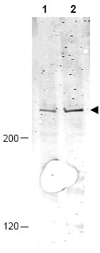
- Western blot using Affinity Purified anti-APC1 pS377 antibody shows detection of a band ~215 kDa corresponding to phosphorylated human APC1 (arrowhead). Lane 1 shows lysate from asynchronous cells. Lane 2 shows lysate from cells treated with nocodazole. While some phosphorylated APC1 is present in untreated cell, the amount of phosphorylated protein is increased in cell preparations arrested in mitosis. Each lane contains approximately 30 µg of HeLa whole cell lysates, separated by 4-8% SDS-PAGE followed by transfer to nitrocellulose. After blocking the membrane was probed with the primary antibody diluted to 1:1,000 overnight at 4°C followed by washes and reaction with a 1:10,000 dilution of IRDye(TM)800 conjugated Gt-a-Rabbit IgG [H&L] MX for 45 min at room temperature. IRDye(TM)800 fluorescence image was captured using the Odyssey(R) Infrared Imaging System developed by LI-COR. IRDye is a trademark of LI-COR, Inc. Other detection systems will yield similar results.
Supportive validation
- Submitted by
- Acris Antibodies GmbH (provider)
- Main image
- Experimental details
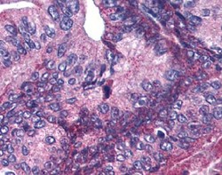
- Immunohistochemistry. Affinity purified anti-APC1 pS377 antibody was used at 5.0 µg/ml to detect signal in a variety of tissues including multi-human, multi-brain and multicancer slides. This image shows moderate positive cytoplasmic and occasional nuclear staining of pancreatic carcinoma cells at 60X. Tissue was formalin-fixed and paraffin embedded. The image shows localization of the antibody as the precipitated red signal, with a hematoxylin purple nuclear counterstain.
