Explore
 Validate
 Learn
Learn Western blot
Antibody data
- Antibody Data
- Antigen structure
- References [2]
- Comments [0]
- Validations
- Western blot [1]
Submit
Validation data
Reference
Comment
Report error
- Product number
- PAB10017 - Provider product page

- Provider
- Abnova Corporation
- Proper citation
- Abnova Corporation Cat#PAB10017, RRID:AB_1676185
- Product name
- ANAPC1 (phospho S355) polyclonal antibody
- Antibody type
- Polyclonal
- Description
- Rabbit polyclonal antibody raised against synthetic phosphopeptide of ANAPC1.
- Storage
- Store at 4°C. For long term storage store at -20°C.Aliquot to avoid repeated freezing and thawing.
Submitted references Mitotic regulation of the human anaphase-promoting complex by phosphorylation.
Characterisation of the human APC1, the largest subunit of the anaphase-promoting complex.
Kraft C, Herzog F, Gieffers C, Mechtler K, Hagting A, Pines J, Peters JM
The EMBO journal 2003 Dec 15;22(24):6598-609
The EMBO journal 2003 Dec 15;22(24):6598-609
Characterisation of the human APC1, the largest subunit of the anaphase-promoting complex.
Jörgensen PM, Gräslund S, Betz R, Ståhl S, Larsson C, Höög C
Gene 2001 Jan 10;262(1-2):51-9
Gene 2001 Jan 10;262(1-2):51-9
No comments: Submit comment
Supportive validation
- Submitted by
- Abnova Corporation (provider)
- Main image
- Experimental details
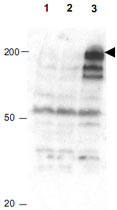
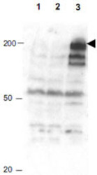
- Western blot using ANAPC1 (phospho S355) polyclonal antibody (Cat # PAB10017) shows detection of a band ~215 KDa corresponding to phosphorylated human ANAPC1 (arrowhead).Lane 1 shows lysate from asynchronous cells.Lane 2 shows lysate from cells treated with thymidine to synchronize cells at the G1/S boundary.Lane 3 shows lysate from cells treated with nocodazole to synchronizecells at the M phase.Phosphorylated ANAPC1 is mostly present only in cell preparations arrested at cell division.Each lane contains approximately 30 ug of HeLa S3 whole cell lysates separated by 12.5% SDS-PAGE followed by transfer to nitrocellulose.After blocking with 5% non-fat drymilk in TTBS, the membrane was probed with the primary antibody diluted to 1 : 500 for 1 h at room temperature followed by washes and reaction with a 1 : 5,000 dilution of HRP Gt-a-Rabbit IgG [H&L] MX for 45 min at room temperature.ECL reagent was used for detection.Data contributed by Bing Li, UT South western.