 Explore
 Validate
 Learn
Learn Western blot
 ELISA
ELISAAntibody data
- Antibody Data
- Antigen structure
- References [0]
- Comments [0]
- Validations
- Western blot [1]
Submit
Validation data
Reference
Comment
Report error
- Product number
- AP09273PU-N - Provider product page

- Provider
- Acris Antibodies GmbH
- Proper citation
- Acris Antibodies GmbH Cat#AP09273PU-N, RRID:AB_2035135
- Product name
- anti APC1 / ANAPC1 pSer355
- Antibody type
- Polyclonal
- Antigen
- Synthetic peptide corresponding to amino acids 351-359 of Human Apc1 protein
- Reactivity
- Human, Mouse, Rat, Bovine, Canine, Chicken/Avian
- Host
- Rabbit
- Isotype
- IgG
- Vial size
- 0.1 mg
- Concentration
- 0.55 mg/ml (by UV absorbance at 280 nm)
No comments: Submit comment
Supportive validation
- Submitted by
- Acris Antibodies GmbH (provider)
- Main image
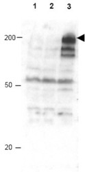
- Experimental details
- Western blot using Affinity Purified anti-APC1 pS355 antibody shows detection of a band ~215 kDa corresponding to phosphorylated human APC1 (arrowhead). Lane 1 shows lysate from asynchronous cells. Lane 2 shows lysate from cells treated with thymidine to synchronize cells at the G1/S boundary. Lane 3 shows lysate from cells treated with nocodazole to synchronize cells at the M phase. Phosphorylated APC1 is mostly present only in cell preparations arrested at cell division. Each lane contains approximately 30 µg of HeLa S3 whole cell lysates separated by 12.5% SDS-PAGE followed by transfer to nitrocellulose. After blocking with 5% non-fat dry milk in TTBS, the membrane was probed with the primary antibody diluted to 1:500 for 1 h at room temperature followed by washes and reaction with a 1:5,000 dilution of HRP Gt-a-Rabbit IgG [H&L] MX for 45 min at room temperature. ECL reagent was used for detection. Other detection systems will yield similar results.
