 Explore
 Validate
 Learn
Learn Immunocytochemistry
Antibody data
- Antibody Data
- Antigen structure
- References [2]
- Comments [0]
- Validations
- Immunocytochemistry [1]
- Other assay [3]
Submit
Validation data
Reference
Comment
Report error
- Product number
- 720107 - Provider product page

- Provider
- Invitrogen Antibodies
- Product name
- Phospho-ATR (Ser428) Polyclonal Antibody
- Antibody type
- Polyclonal
- Antigen
- Synthetic peptide
- Description
- These Polyclonal antibodies are of rabbit origin developed by immunizing animals with proteins or peptides. The polyclonal antibody is purified by affinity purification from the rabbit sera generated after immunizing the rabbits with a specific type of protein or peptide. The purified antibody is tested for its functionality in various relevant research applications. The antibody is developed for Research Use Only and is non-hazardous or non-infectious in nature.
- Concentration
- 0.5 mg/mL
Submitted references Cyclin F is involved in response to cisplatin treatment in melanoma cell lines.
The ATM and Rad3-Related (ATR) Protein Kinase Pathway Is Activated by Herpes Simplex Virus 1 and Required for Efficient Viral Replication.
Krajewski A, Gagat M, Żuryń A, Hałas-Wiśniewska M, Grzanka D, Grzanka A
Oncology reports 2020 Mar;43(3):765-772
Oncology reports 2020 Mar;43(3):765-772
The ATM and Rad3-Related (ATR) Protein Kinase Pathway Is Activated by Herpes Simplex Virus 1 and Required for Efficient Viral Replication.
Edwards TG, Bloom DC, Fisher C
Journal of virology 2018 Mar 15;92(6)
Journal of virology 2018 Mar 15;92(6)
No comments: Submit comment
Supportive validation
- Submitted by
- Invitrogen Antibodies (provider)
- Main image

- Experimental details
- For immunofluorescence analysis HeLa cells were fixed and permeabilized for detection of ATR (pS428) using ATR (pS428) Rabbit Polyclonal antibody (Product # 720107, 2 µg/mL) and labeled with Goat anti-Rabbit IgG (H+L) Superclonal™ Secondary Antibody, Alexa Fluor® 488 conjugate (Product # A27034, 1:2000). Nuclei (blue) were stained using SlowFade® Gold Antifade Mountant with DAPI (Product # S36938), and Alexa Fluor® 555 Rhodamine Phalloidin (Product # R415, 1:300) was used for cytoskeletal F-actin (red) staining. Detection and localization of ATR (pS428) (green) in the nucleus can be clearly observed in cells treated with hydroxyurea (HU, 3 mM, 20 h) as compared to untreated cells. Antibody specificity was demonstrated by competition with the ATR (pS428) peptide, which results in complete loss of signal. No competition was observed with the non-phospho peptide.

Supportive validation
- Submitted by
- Invitrogen Antibodies (provider)
- Main image
- Experimental details
- NULL

- Submitted by
- Invitrogen Antibodies (provider)
- Main image
- Experimental details
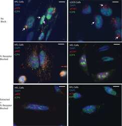
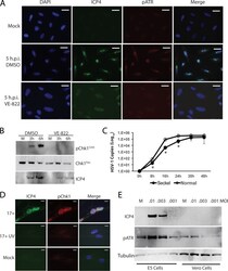
- FIG 6 The ATR kinase inhibitor VE-822 blocks activation of ATR and Chk1, while HSV-1 replication is deficient in Seckel patient fibroblasts hypomorphic for ATR. (A) Immunofluorescence images of ICP4 and pATR at 5 h.p.i. with HSV-1 17 syn + (MOI = 10) and treatment of U2OS cells with either a vehicle (DMSO) or VE-822 (10 muM). Cells are unextracted. Scale bars = 20 mum. (B) Western blot demonstrating that VE-822 (10 muM) also blocks phosphorylation of Chk1 in HSV-1-infected U2OS cells while suppressing ICP4 expression. (C) Seckel syndrome patient fibroblasts, hypomorphic for ATR, are defective for HSV-1 replication relative to control patient (IBR3) fibroblasts. (D) UV-inactivated HSV-1 17 syn + (17+ UV; MOI = 10) does not significantly activate the ATR pathway. Cells are unextracted. Scale bars = 10 mum. (E) An ICP4 - , replication-incompetent mutant HSV-1 virus (KD6) does not express ICP4 and does not activate the ATR pathway in nonpermissive (Vero) cells following 24 h of infection with a low MOI. The permissive cell line E5, which supplies ICP4, shows expression of ICP4 and activation of the ATR pathway.

- Submitted by
- Invitrogen Antibodies (provider)
- Main image
- Experimental details
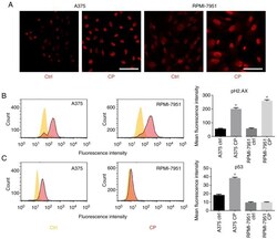
- Figure 3. (A) Immunofluorescence staining of p-ATR in A375 and RPMI-7951 cell lines. (B) Flow cytometric analysis of p-H2.AX expression in A375 and RPMI-7951 cell lines. (C) Flow cytometric analysis of p53 expression in A375 and RPMI-7951 cell lines. *P
