Explore
 Validate
 Learn
Learn Western blot
 Immunocytochemistry
Antibody data
- Antibody Data
- Antigen structure
- References [20]
- Comments [0]
- Validations
- Immunocytochemistry [1]
- Chromatin Immunoprecipitation [1]
Submit
Validation data
Reference
Comment
Report error
- Product number
- HPA006111 - Provider product page

- Provider
- Atlas Antibodies
- Proper citation
- Atlas Antibodies Cat#HPA006111, RRID:AB_1854422
- Product name
- Anti-NFIA
- Antibody type
- Polyclonal
- Description
- Polyclonal Antibody against Human NFIA, Gene description: nuclear factor I/A, Alternative Gene Names: KIAA1439, NFI-L, Validated applications: ChIP, ICC, IHC, WB, Uniprot ID: Q12857, Storage: Store at +4°C for short term storage. Long time storage is recommended at -20°C.
- Reactivity
- Human, Mouse, Rat
- Host
- Rabbit
- Conjugate
- Unconjugated
- Isotype
- IgG
- Vial size
- 100 µl
- Concentration
- 0.1 mg/ml
- Storage
- Store at +4°C for short term storage. Long time storage is recommended at -20°C.
- Handling
- The antibody solution should be gently mixed before use.
Submitted references Systemic and intrinsic functions of ATRX in glial cell fate and CNS myelination in male mice
NFIA in adipocytes reciprocally regulates mitochondrial and inflammatory gene program to improve glucose homeostasis
Human Alzheimer’s disease reactive astrocytes exhibit a loss of homeostastic gene expression
Injury primes mutation-bearing astrocytes for dedifferentiation in later life.
Identification and validation of NFIA as a novel prognostic marker in renal cell carcinoma.
Lunatic Fringe-GFP Marks Lamina-Specific Astrocytes That Regulate Sensory Processing
NFIA determines the cis-effect of genetic variation on Ucp1 expression in murine thermogenic adipocytes
Nuclear Factor I in neurons, glia and during the formation of Müller glia-derived progenitor cells in avian, porcine and primate retinas.
Mapping Astrocyte Transcriptional Signatures in Response to Neuroactive Compounds
Capicua regulates the development of adult-born neurons in the hippocampus
NFIA differentially controls adipogenic and myogenic gene program through distinct pathways to ensure brown and beige adipocyte differentiation
Region-Specific Transcriptional Control of Astrocyte Function Oversees Local Circuit Activities
Cep215 is essential for morphological differentiation of astrocytes
Nuclear factor I-A regulates diverse reactive astrocyte responses after CNS injury
Sequentially acting SOX proteins orchestrate astrocyte‐ and oligodendrocyte‐specific gene expression
DNER and NFIA are expressed by developing and mature AII amacrine cells in the mouse retina.
Chd7 Collaborates with Sox2 to Regulate Activation of Oligodendrocyte Precursor Cells after Spinal Cord Injury
Zbtb20 promotes astrocytogenesis during neocortical development
Rowland M, Jiang Y, Shafiq S, Ghahramani A, Pena-Ortiz M, Dumeaux V, Bérubé N
Nature Communications 2023;14(1)
Nature Communications 2023;14(1)
NFIA in adipocytes reciprocally regulates mitochondrial and inflammatory gene program to improve glucose homeostasis
Hiraike Y, Saito K, Oguchi M, Wada T, Toda G, Tsutsumi S, Bando K, Sagawa J, Nagano G, Ohno H, Kubota N, Kubota T, Aburatani H, Kadowaki T, Waki H, Yanagimoto S, Yamauchi T
Proceedings of the National Academy of Sciences 2023;120(31)
Proceedings of the National Academy of Sciences 2023;120(31)
Human Alzheimer’s disease reactive astrocytes exhibit a loss of homeostastic gene expression
Dai D, Li M, Lee E
Acta Neuropathologica Communications 2023;11(1)
Acta Neuropathologica Communications 2023;11(1)
Injury primes mutation-bearing astrocytes for dedifferentiation in later life.
Simpson Ragdale H, Clements M, Tang W, Deltcheva E, Andreassi C, Lai AG, Chang WH, Pandrea M, Andrew I, Game L, Uddin I, Ellis M, Enver T, Riccio A, Marguerat S, Parrinello S
Current biology : CB 2023 Mar 27;33(6):1082-1098.e8
Current biology : CB 2023 Mar 27;33(6):1082-1098.e8
Identification and validation of NFIA as a novel prognostic marker in renal cell carcinoma.
de Alwis R, Schoch S, Islam M, Möller C, Ljungberg B, Axelson H
The journal of pathology. Clinical research 2023 Jul;9(4):261-272
The journal of pathology. Clinical research 2023 Jul;9(4):261-272
Scavuzzo M, Letai K, Maeno-Hikichi Y, Wulftange W, Shah I, Rameshbabu J, Tomar A, Shick H, Shah A, Xiong Y, Cohn E, Allan K, Tesar P
2023
2023
Cheng Y, Luna-Figueroa E, Woo J, Chen H, Lee Z, Harmanci A, Deneen B
2023
2023
Lunatic Fringe-GFP Marks Lamina-Specific Astrocytes That Regulate Sensory Processing
Akdemir E, Woo J, Bosquez Huerta N, Lozzi B, Groves A, Harmanci A, Deneen B
The Journal of Neuroscience 2022;42(4):567-580
The Journal of Neuroscience 2022;42(4):567-580
NFIA determines the cis-effect of genetic variation on Ucp1 expression in murine thermogenic adipocytes
Hiraike Y, Tsutsumi S, Wada T, Oguchi M, Saito K, Nakamura M, Ota S, Koebis M, Nakao H, Aiba A, Nagano G, Ohno H, Oki K, Yoneda M, Kadowaki T, Aburatani H, Waki H, Yamauchi T
iScience 2022;25(8):104729
iScience 2022;25(8):104729
Nuclear Factor I in neurons, glia and during the formation of Müller glia-derived progenitor cells in avian, porcine and primate retinas.
El-Hodiri HM, Campbell WA, Kelly LE, Hawthorn EC, Schwartz M, Jalligampala A, McCall MA, Meyer K, Fischer AJ
The Journal of comparative neurology 2022 Jun;530(8):1213-1230
The Journal of comparative neurology 2022 Jun;530(8):1213-1230
Mapping Astrocyte Transcriptional Signatures in Response to Neuroactive Compounds
Sardar D, Lozzi B, Woo J, Huang T, Cvetkovic C, Creighton C, Krencik R, Deneen B
International Journal of Molecular Sciences 2021;22(8):3975
International Journal of Molecular Sciences 2021;22(8):3975
Capicua regulates the development of adult-born neurons in the hippocampus
Hourigan B, Balay S, Yee G, Sharma S, Tan Q
Scientific Reports 2021;11(1)
Scientific Reports 2021;11(1)
NFIA differentially controls adipogenic and myogenic gene program through distinct pathways to ensure brown and beige adipocyte differentiation
Conlon F, Hiraike Y, Waki H, Miyake K, Wada T, Oguchi M, Saito K, Tsutsumi S, Aburatani H, Yamauchi T, Kadowaki T
PLOS Genetics 2020;16(9):e1009044
PLOS Genetics 2020;16(9):e1009044
Region-Specific Transcriptional Control of Astrocyte Function Oversees Local Circuit Activities
Huang A, Woo J, Sardar D, Lozzi B, Bosquez Huerta N, Lin C, Felice D, Jain A, Paulucci-Holthauzen A, Deneen B
Neuron 2020;106(6):992-1008.e9
Neuron 2020;106(6):992-1008.e9
Cep215 is essential for morphological differentiation of astrocytes
Kang D, Shin W, Yoo H, Kim S, Lee S, Rhee K
Scientific Reports 2020;10(1)
Scientific Reports 2020;10(1)
Nuclear factor I-A regulates diverse reactive astrocyte responses after CNS injury
Laug D, Huang T, Huerta N, Huang A, Sardar D, Ortiz-Guzman J, Carlson J, Arenkiel B, Kuo C, Mohila C, Glasgow S, Lee H, Deneen B
Journal of Clinical Investigation 2019;129(10):4408-4418
Journal of Clinical Investigation 2019;129(10):4408-4418
Sequentially acting SOX proteins orchestrate astrocyte‐ and oligodendrocyte‐specific gene expression
Klum S, Zaouter C, Alekseenko Z, Björklund Å, Hagey D, Ericson J, Muhr J, Bergsland M
EMBO reports 2018;19(11)
EMBO reports 2018;19(11)
DNER and NFIA are expressed by developing and mature AII amacrine cells in the mouse retina.
Keeley PW, Reese BE
The Journal of comparative neurology 2018 Feb 15;526(3):467-479
The Journal of comparative neurology 2018 Feb 15;526(3):467-479
Chd7 Collaborates with Sox2 to Regulate Activation of Oligodendrocyte Precursor Cells after Spinal Cord Injury
Doi T, Ogata T, Yamauchi J, Sawada Y, Tanaka S, Nagao M
The Journal of Neuroscience 2017;37(43):10290-10309
The Journal of Neuroscience 2017;37(43):10290-10309
Zbtb20 promotes astrocytogenesis during neocortical development
Nagao M, Ogata T, Sawada Y, Gotoh Y
Nature Communications 2016;7(1)
Nature Communications 2016;7(1)
No comments: Submit comment
Supportive validation
- Submitted by
- Atlas Antibodies (provider)
- Main image
- Experimental details
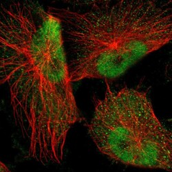
- Immunofluorescent staining of human cell line U-251 MG shows localization to nucleoplasm.
- Sample type
- Human

Supportive validation
- Submitted by
- Atlas Antibodies (provider)
- Main image
- Experimental details
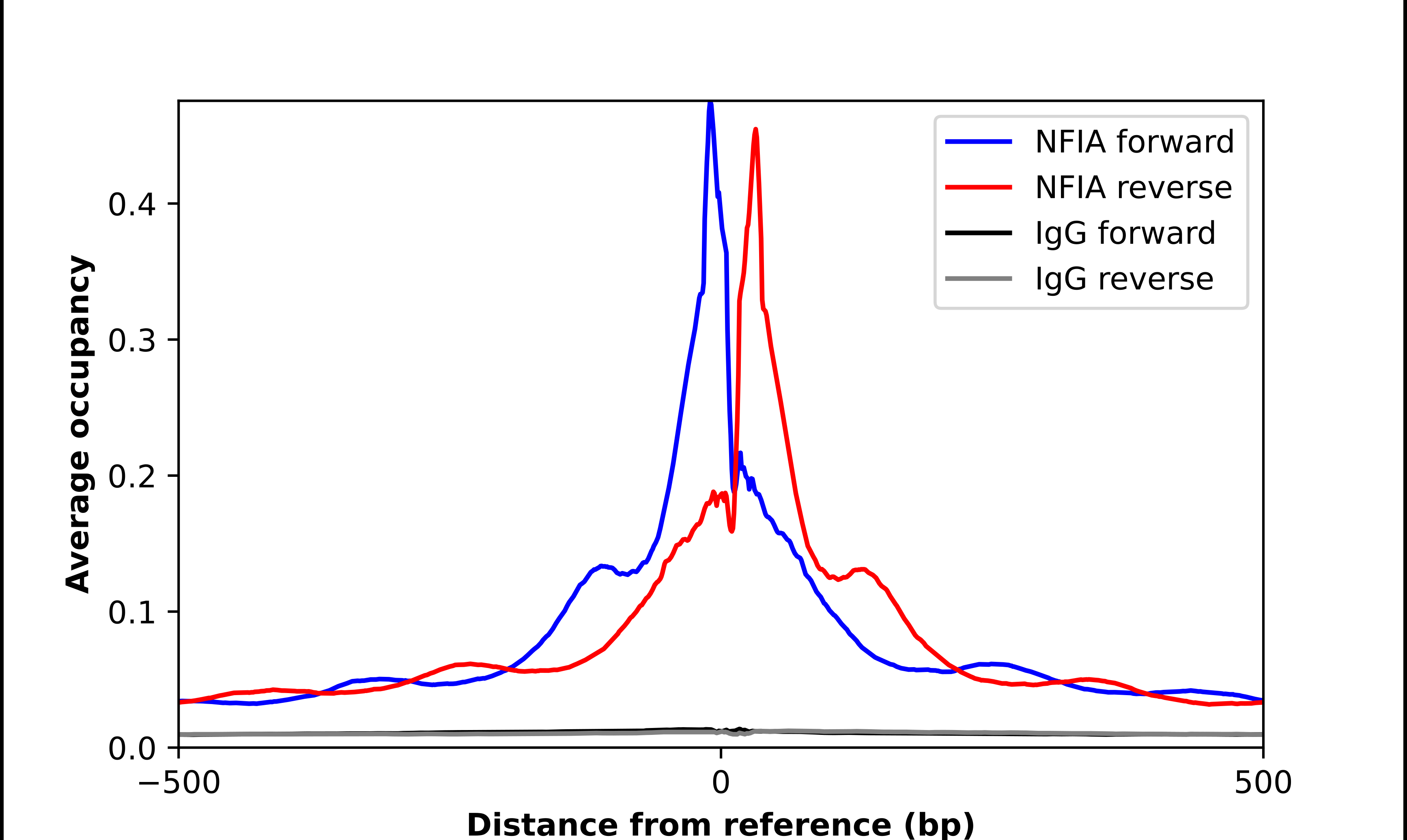
- ChIP-Exo-Seq composite graph for Anti-NFIA (HPA006111 , Lot 000007218) tested in K562 cells. Strand-specific reads (blue: forward, red: reverse) and IgG controls (black: forward, grey: reverse) are plotted against the distance from a composite set of reference binding sites. The antibody exhibits robust target enrichment compared to a non-specific IgG control and precisely reveals its structural organization around the binding site. Data generated by Prof. B. F. Pugh´s Lab at Cornell University.