Explore
 Validate
 Learn
Learn Western blot
Antibody data
- Antibody Data
- Antigen structure
- References [0]
- Comments [0]
- Validations
- Western blot [4]
- Immunocytochemistry [1]
- Immunohistochemistry [1]
Submit
Validation data
Reference
Comment
Report error
- Product number
- PA5-21649 - Provider product page

- Provider
- Invitrogen Antibodies
- Product name
- PLSCR1 Polyclonal Antibody
- Antibody type
- Polyclonal
- Antigen
- Recombinant protein fragment
- Description
- Recommended positive controls: 293T, A431, H1299, HeLaS3, HepG2, Molt-4. Predicted reactivity: Rhesus Monkey (96%). Store product as a concentrated solution. Centrifuge briefly prior to opening the vial.
- Reactivity
- Human
- Host
- Rabbit
- Isotype
- IgG
- Vial size
- 100 µL
- Concentration
- 0.52 mg/mL
- Storage
- Store at 4°C short term. For long term storage, store at -20°C, avoiding freeze/thaw cycles.
No comments: Submit comment
Supportive validation
- Submitted by
- Invitrogen Antibodies (provider)
- Main image

- Experimental details
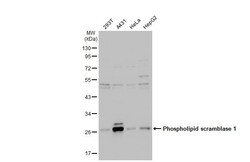
- Western blot analysis of phospholipid scramblase 1 using 30 µg of HeLa S3 lysate. Samples were loaded onto a 10% SDS-PAGE gel and probed with a phospholipid scramblase 1 polyclonal antibody (Product # PA5-21649) at a dilution of 1:1000.

- Submitted by
- Invitrogen Antibodies (provider)
- Main image
- Experimental details
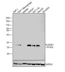
- Western blot was performed using Anti-PLSCR1 Polyclonal Antibody (Product # PA5-21649) and a 35 kDa band corresponding to Phospholipid scramblase 1 was observed across positive cell lines (THP1 (low expressing), THP1 Macrophage, HaCaT, HCT 116 and Hep G2); and not across negative models (SW480 and SH-SY5Y). Membrane enriched extracts (30 µg lysate) of THP-1 (Lane 1), THP-1 Macrophage (Lane 2), SW480 (Lane 3), HaCaT (Lane 4), HCT 116 (Lane 5), Hep G2 (Lane 6) and SH-SY5Y (Lane 7) were electrophoresed using NuPAGE™ 10% Bis-Tris Protein Gel (Product # NP0301BOX). Resolved proteins were then transferred onto a nitrocellulose membrane (Product # IB23001) by iBlot® 2 Dry Blotting System (Product # IB21001). The blot was probed with the primary antibody (1:1000 dilution) and detected by chemiluminescence with Goat anti-Rabbit IgG (H+L) Superclonal™ Recombinant Secondary Antibody, HRP (Product # A27036,1:20000 dilution) using the iBright™ FL1500 Imaging System (Product # A44115). Chemiluminescent detection was performed using SuperSignal™ West Atto Ultimate Sensitivity Substrate (Product # A38556).

- Submitted by
- Invitrogen Antibodies (provider)
- Main image
- Experimental details
- Western Blot using PLSCR1 Polyclonal Antibody (Product # PA5-21649). Various whole cell extracts (30 µg) were separated by 10% SDS-PAGE, and the membrane was blotted with PLSCR1 Polyclonal Antibody (Product # PA5-21649) diluted at 1:1,000. The HRP-conjugated anti-rabbit IgG antibody was used to detect the primary antibody.

- Submitted by
- Invitrogen Antibodies (provider)
- Main image

- Experimental details
- Western blot was performed using Anti-PLSCR1 Polyclonal Antibody (Product # PA5-21649) and a 35 kDa band corresponding to Phospholipid scramblase 1 was observed across positive cell lines (THP1 (low expressing), THP1 Macrophage, HaCaT, HCT 116 and Hep G2); and not across negative models (SW480 and SH-SY5Y). Membrane enriched extracts (30 µg lysate) of THP-1 (Lane 1), THP-1 Macrophage (Lane 2), SW480 (Lane 3), HaCaT (Lane 4), HCT 116 (Lane 5), Hep G2 (Lane 6) and SH-SY5Y (Lane 7) were electrophoresed using NuPAGE™ 10% Bis-Tris Protein Gel (Product # NP0301BOX). Resolved proteins were then transferred onto a nitrocellulose membrane (Product # IB23001) by iBlot® 2 Dry Blotting System (Product # IB21001). The blot was probed with the primary antibody (1:1000 dilution) and detected by chemiluminescence with Goat anti-Rabbit IgG (H+L) Superclonal™ Recombinant Secondary Antibody, HRP (Product # A27036,1:20000 dilution) using the iBright™ FL1500 Imaging System (Product # A44115). Chemiluminescent detection was performed using SuperSignal™ West Atto Ultimate Sensitivity Substrate (Product # A38556).

Supportive validation
- Submitted by
- Invitrogen Antibodies (provider)
- Main image
- Experimental details
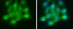
- Immunocytochemistry-Immunofluorescence analysis of PLSCR1 was performed in A431 cells fixed in ice-cold MeOH for 5 min. Green: PLSCR1 Polyclonal Antibody (Product # PA5-21649) diluted at 1:5000. Blue: Hoechst 33342 staining.

Supportive validation
- Submitted by
- Invitrogen Antibodies (provider)
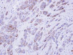
- Main image
- Experimental details
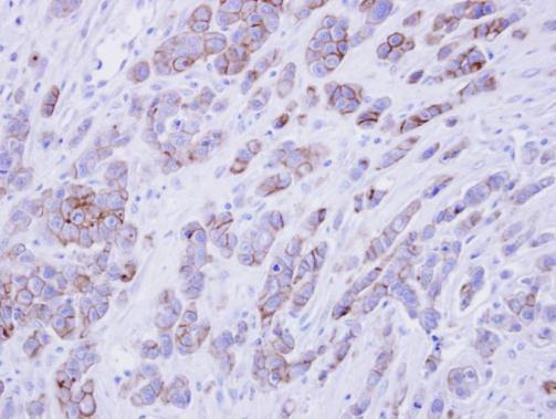
- Immunohistochemical analysis of paraffin-embedded MDAMB468 xenograft , using phospholipid scramblase 1 (Product # PA5-21649) antibody at 1:500 dilution. Antigen Retrieval: EDTA based buffer, pH 8.0, 15 min.