Explore
 Validate
 Learn
Learn Western blot
 Immunohistochemistry
Antibody data
- Antibody Data
- Antigen structure
- References [4]
- Comments [0]
- Validations
- Immunohistochemistry [1]
Submit
Validation data
Reference
Comment
Report error
- Product number
- HPA043673 - Provider product page

- Provider
- Atlas Antibodies
- Proper citation
- Atlas Antibodies Cat#HPA043673, RRID:AB_10960893
- Product name
- Anti-COL17A1
- Antibody type
- Polyclonal
- Description
- Polyclonal Antibody against Human COL17A1, Gene description: collagen, type XVII, alpha 1, Alternative Gene Names: BP180, BPAG2, Validated applications: WB, IHC, Uniprot ID: Q9UMD9, Storage: Store at +4°C for short term storage. Long time storage is recommended at -20°C.
- Reactivity
- Human
- Host
- Rabbit
- Conjugate
- Unconjugated
- Isotype
- IgG
- Vial size
- 100 µl
- Concentration
- 0.3 mg/ml
- Storage
- Store at +4°C for short term storage. Long time storage is recommended at -20°C.
- Handling
- The antibody solution should be gently mixed before use.
Submitted references Functional analysis of Collagen 17a1: A genetic modifier of junctional epidermolysis bullosa in mice.
Multimodal Analysis of Composition and Spatial Architecture in Human Squamous Cell Carcinoma
Dual roles of hemidesmosomal proteins in the pancreatic epithelium: the phosphoinositide 3-kinase decides
Sproule TJ, Wilpan RY, Low BE, Silva KA, Reyon D, Joung JK, Wiles MV, Roopenian DC, Sundberg JP
PloS one 2023;18(10):e0292456
PloS one 2023;18(10):e0292456
Jackson R, Rajadhyaksha E, Loeffler R, Flores C, Van Doorslaer K
2023
2023
Multimodal Analysis of Composition and Spatial Architecture in Human Squamous Cell Carcinoma
Ji A, Rubin A, Thrane K, Jiang S, Reynolds D, Meyers R, Guo M, George B, Mollbrink A, Bergenstråhle J, Larsson L, Bai Y, Zhu B, Bhaduri A, Meyers J, Rovira-Clavé X, Hollmig S, Aasi S, Nolan G, Lundeberg J, Khavari P
Cell 2020;182(2):497-514.e22
Cell 2020;182(2):497-514.e22
Dual roles of hemidesmosomal proteins in the pancreatic epithelium: the phosphoinositide 3-kinase decides
Laval S, Laklai H, Fanjul M, Pucelle M, Laurell H, Billon-Galés A, Le Guellec S, Delisle M, Sonnenberg A, Susini C, Pyronnet S, Bousquet C
Oncogene 2013;33(15):1934-1944
Oncogene 2013;33(15):1934-1944
No comments: Submit comment
Supportive validation
- Submitted by
- Atlas Antibodies (provider)
- Enhanced method
- Orthogonal validation
- Main image
- Experimental details
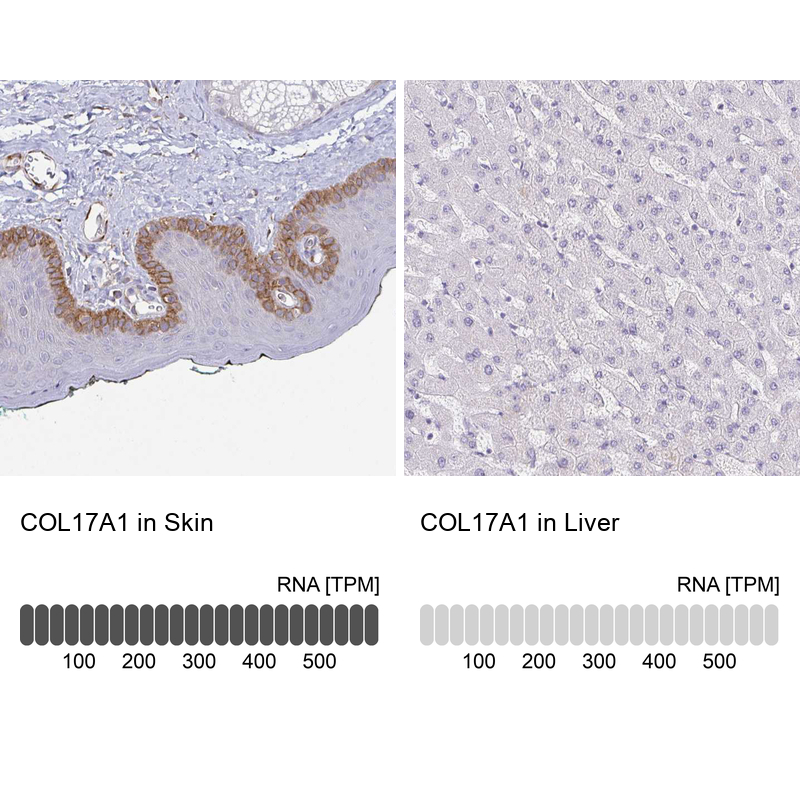
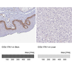
- Immunohistochemistry analysis in human skin and liver tissues using HPA043673 antibody. Corresponding COL17A1 RNA-seq data are presented for the same tissues.
- Sample type
- Human
- Protocol
- Protocol