Explore
 Validate
 Learn
Learn Western blot
Antibody data
- Antibody Data
- Antigen structure
- References [1]
- Comments [0]
- Validations
- Western blot [3]
- Immunocytochemistry [1]
- Immunohistochemistry [2]
- Flow cytometry [2]
Submit
Validation data
Reference
Comment
Report error
- Product number
- TA501358 - Provider product page

- Provider
- OriGene
- Proper citation
- OriGene Cat#TA501358, RRID:AB_11126863
- Product name
- Anti-TBXAS1 (Thromboxane synthase) mouse monoclonal antibody, clone OTI2C1 (formerly 2C1)
- Antibody type
- Monoclonal
- Description
- Anti-TBXAS1 (Thromboxane synthase) mouse monoclonal antibody, clone OTI2C1 (formerly 2C1)
- Host
- Mouse
- Conjugate
- Unconjugated
- Epitope
- TBXAS1
- Isotype
- IgG
- Antibody clone number
- OTI2C1
- Vial size
- 100 µl
- Concentration
- 0.86 mg/ml
Submitted references Glycyrrhizin suppresses lung adenocarcinoma cell growth through inhibition of thromboxane synthase.
Huang RY, Chu YL, Jiang ZB, Chen XM, Zhang X, Zeng X
Cellular physiology and biochemistry : international journal of experimental cellular physiology, biochemistry, and pharmacology 2014;33(2):375-88
Cellular physiology and biochemistry : international journal of experimental cellular physiology, biochemistry, and pharmacology 2014;33(2):375-88
No comments: Submit comment
Supportive validation
- Submitted by
- OriGene (provider)
- Main image
- Experimental details
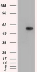
- HEK293T cells were transfected with the pCMV6-ENTRY control (Left lane) or pCMV6-ENTRY TBXAS1 (RC208028, Right lane) cDNA for 48 hrs and lysed. Equivalent amounts of cell lysates (5 ug per lane) were separated by SDS-PAGE and immunoblotted with anti-TBXAS1.
- Validation comment
- WB
- Submitted by
- OriGene (provider)
- Main image
- Experimental details
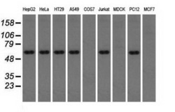
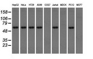
- Western blot analysis of extracts (35ug) from 9 different cell lines by using anti-TBXAS1 monoclonal antibody.
- Validation comment
- WB
- Submitted by
- OriGene (provider)
- Main image
- Experimental details
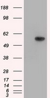
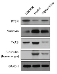
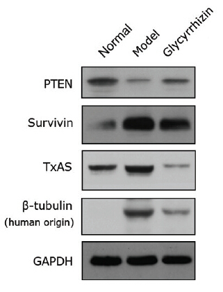
- Figure from citation: Western Blot of TBXAS1(TxAS) protein level by using anti-TBXAS1 antibody in human lung adenocarcinoma. Dilution: 1:1000
- Validation comment
- WB
Supportive validation
- Submitted by
- OriGene (provider)
- Main image

- Experimental details
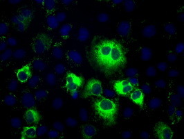
- Anti-TBXAS1 mouse monoclonal antibody (TA501358) immunofluorescent staining of COS7 cells transiently transfected by pCMV6-ENTRY TBXAS1(RC208028).
- Validation comment
- IF
Supportive validation
- Submitted by
- OriGene (provider)
- Main image
- Experimental details
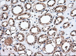
- Immunohistochemical staining of paraffin-embedded Human Kidney tissue within the normal limits using anti-TBXAS1 mouse monoclonal antibody. (Heat-induced epitope retrieval by 10mM citric buffer, pH6.0, 100C for 10min, TA501358, Dilution 1:50)
- Validation comment
- IHC

- Submitted by
- OriGene (provider)
- Main image
- Experimental details
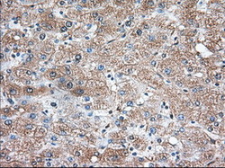
- Immunohistochemical staining of paraffin-embedded Human liver tissue within the normal limits using anti-TBXAS1 mouse monoclonal antibody. (Heat-induced epitope retrieval by 10mM citric buffer, pH6.0, 100C for 10min, TA501358, Dilution 1:50)
- Validation comment
- IHC

Supportive validation
- Submitted by
- OriGene (provider)
- Main image

- Experimental details
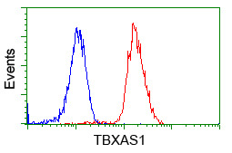
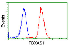
- HEK293T cells transfected with either RC208028 overexpress plasmid(Red) or empty vector control plasmid(Blue) were immunostained by anti-TBXAS1 antibody(TA501358), and then analyzed by flow cytometry.
- Validation comment
- FC
- Submitted by
- OriGene (provider)
- Main image
- Experimental details
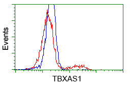
- Flow cytometric Analysis of Jurkat cells, using anti-TBXAS1 antibody(TA501358),(Red), compared to a nonspecific negative control antibody(TA50011),(Blue).
- Validation comment
- FC