Explore
 Validate
 Learn
LearnPA1-41208
antibody from Invitrogen Antibodies
Targeting: SIK2
DKFZp434K1115, KIAA0781, LOH11CR1I, QIK, SNF1LK2
 Western blot
Antibody data
- Antibody Data
- Antigen structure
- References [1]
- Comments [0]
- Validations
- Western blot [1]
- Other assay [2]
Submit
Validation data
Reference
Comment
Report error
- Product number
- PA1-41208 - Provider product page

- Provider
- Invitrogen Antibodies
- Product name
- SIK2 Polyclonal Antibody
- Antibody type
- Polyclonal
- Antigen
- Other
- Description
- Suggested positive control: human brain tissue, human brain protein.
- Reactivity
- Human, Mouse, Rat, Bovine, Canine
- Host
- Rabbit
- Isotype
- IgG
- Vial size
- 100 μg
- Concentration
- 1 mg/mL
- Storage
- Store at 4°C short term. For long term storage, store at -20°C, avoiding freeze/thaw cycles.
Submitted references Role of salt‑inducible kinase 2 in the malignant behavior and glycolysis of colorectal cancer cells.
Ni X, Feng Y, Fu X
Molecular medicine reports 2021 Nov;24(5)
Molecular medicine reports 2021 Nov;24(5)
No comments: Submit comment
Supportive validation
- Submitted by
- Invitrogen Antibodies (provider)
- Main image
- Experimental details
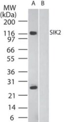
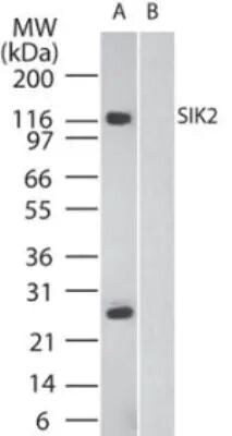
- Western blot analysis of SIK2 in human brain in the A) absence and B) presence of immunizing peptide. Samples were incubated in SIK2 polyclonal antibody (Product # PA1-41208) using a dilution of 2 µg/mL.
Supportive validation
- Submitted by
- Invitrogen Antibodies (provider)
- Main image
- Experimental details
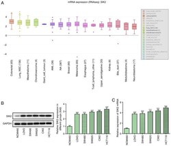
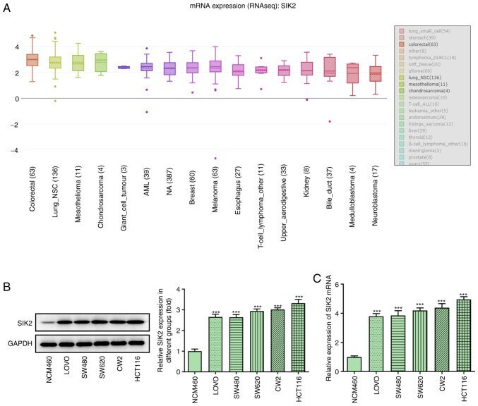
- Figure 1. Expression level of SIK2 in CRC lines. (A) mRNA expression levels of SIK2 were notably increased in all types of cancer in the Cancer Cell Line Encyclopedia. The expression levels of SIK2 in the normal colonic epithelial cell line, NCM460, and different CRC cell lines (LoVo, SW480, SW620, CW2 and HCT116) were determined via (B) western blotting and (C) reverse transcription-quantitative PCR. ***P
- Submitted by
- Invitrogen Antibodies (provider)
- Main image

- Experimental details
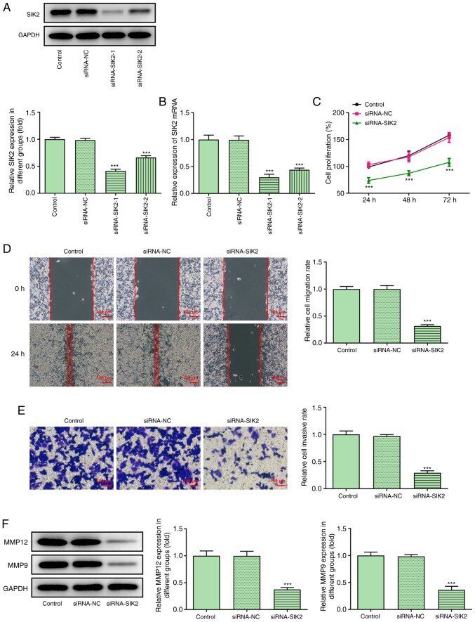
- Figure 2. SIK2 silencing attenuates the proliferation, migration and invasion of colorectal cancer cells. The transfection efficiency in HCT116 cells transfected with siRNA-SIK2 was evaluated via (A) western blot and (B) reverse transcription-quantitative PCR analyses. (C) A Cell Counting Kit-8 assay was used to determine the proliferative ability of HCT116 cells. Cell migration and invasion rates were measured using (D) wound healing and (E) Transwell assays, respectively. Scale bar, 100 um. (F) MMP12 and MMP9 protein expression levels were detected using western blot analysis. ***P