 Explore
 Validate
 Learn
Learn Western blot
Antibody data
- Antibody Data
- Antigen structure
- References [0]
- Comments [0]
- Validations
- Western blot [7]
- Immunocytochemistry [1]
- Immunohistochemistry [1]
- Chromatin Immunoprecipitation [1]
Submit
Validation data
Reference
Comment
Report error
- Product number
- PA5-36279 - Provider product page

- Provider
- Invitrogen Antibodies
- Product name
- NPM1 Polyclonal Antibody
- Antibody type
- Polyclonal
- Antigen
- Synthetic peptide
- Description
- This antibody detects endogenous protein at a molecular weight of 36 kDa.
- Concentration
- 1 mg/mL
No comments: Submit comment
Supportive validation
- Submitted by
- Invitrogen Antibodies (provider)
- Main image

- Experimental details
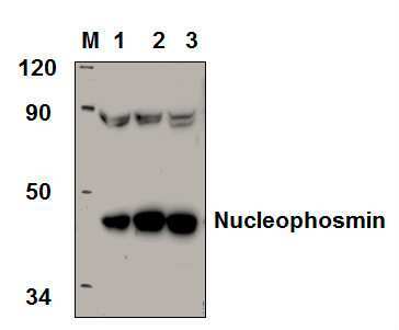
- Western blot analysis of NPM1 in Lane 1: HCT-116 whole cell lysate (40 µg), Lane 2: SGC-7901 whole cell lysate (40 µg), Lane 3: CT-26 whole cell lysate (40 µg). Samples were incubated with NPM1 polyclonal antibody (Product # PA5-36279) at a dilution of 1:1,000.
- Submitted by
- Invitrogen Antibodies (provider)
- Main image

- Experimental details
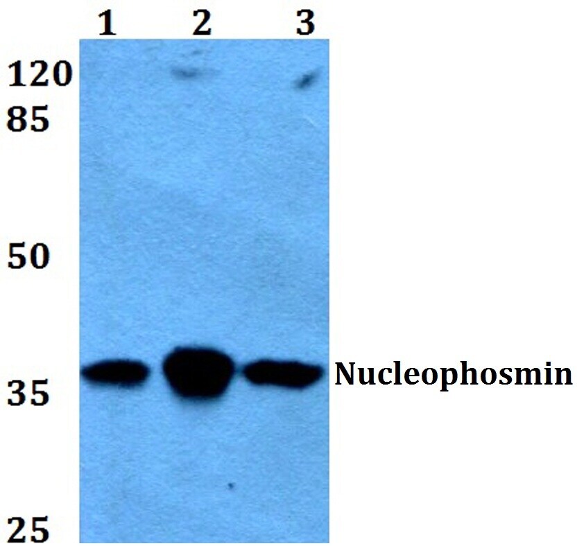
- Western blot analysis of NPM1 in Lane 1: HEK293T whole cell lysate, Lane 2: Raw264.7 whole cell lysate, Lane 3: H9C2 whole cell lysate. Samples were incubated with NPM1 polyclonal antibody (Product # PA5-36279) at a dilution of 1:500.
- Submitted by
- Invitrogen Antibodies (provider)
- Main image

- Experimental details
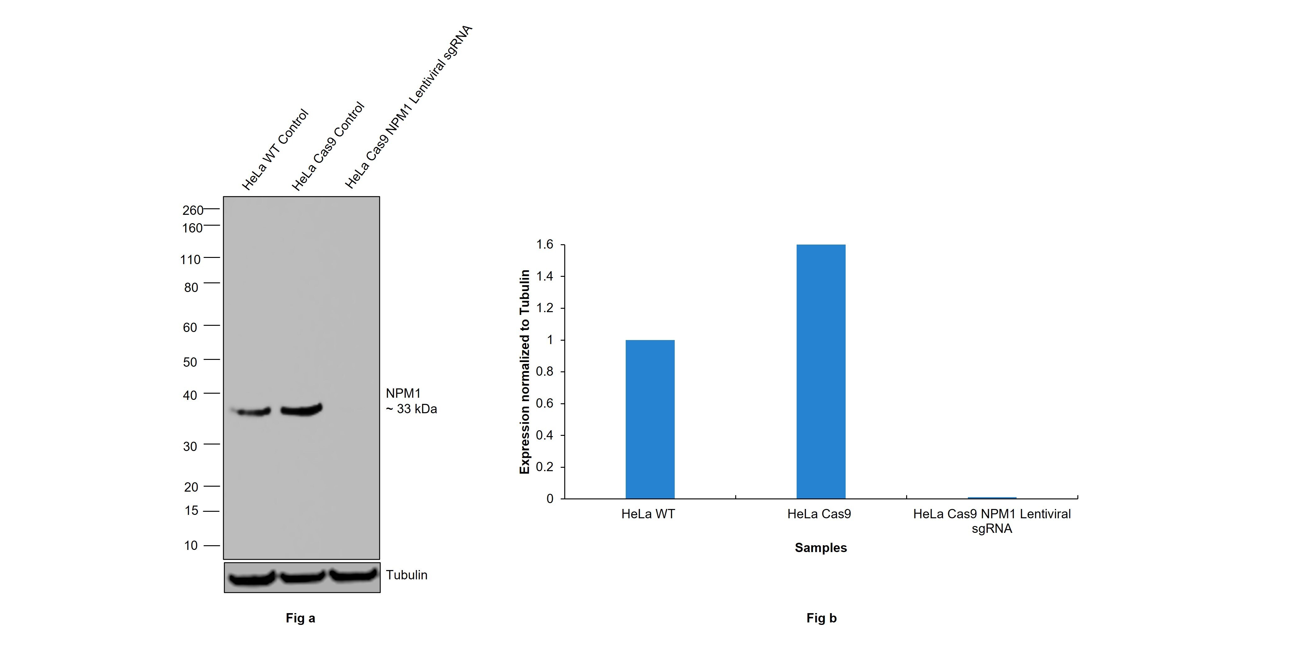
- CRISPR-Cas9 mediated genome editing ofNPM1 (as confirmed by next generation sequencing) was achieved by using LentiArray™ Lentiviral sgRNA (Product # A32042, AssayID CRISPR759267_LV) and LentiArray Cas9 Lentivirus (Product # A32064). Fig (a) Western blot analysis of NPM1 was performed by loading 30 µg of HeLa wild type (Lane 1), HeLa Cas9 (Lane 2) and HeLa Cas9 cells transduced with NPM1 Lentiviral sgRNA (Lane 3) modified whole cell extracts. The samples were electrophoresed using NuPAGE™ Novex™ 4-12% Bis-Tris Protein Gel (Product # NP0322BOX). Resolved proteins were then transferred onto a nitrocellulose membrane (Product # IB23001) by iBlot® 2 Dry Blotting System (Product # IB21001). The blot was probed with NPM1 Polyclonal Antibody (Product # PA5-36279, 1:1200 dilution) and Goat anti-Rabbit IgG (H+L) Superclonal™ Recombinant Secondary Antibody, HRP (Product # A27036 1:10,000 dilution). Chemiluminescent detection was performed using SuperSignal™ West Dura Extended Duration Substrate (Product # 34076). A loss of signal in sgRNA transduced cells using the LentiArray™ CRISPR product line confirms that antibody is specific toNPM1 (Fig (b)).
- Submitted by
- Invitrogen Antibodies (provider)
- Main image

- Experimental details
- Western blot analysis of NPM1 in Lane 1: HCT-116 whole cell lysate (40 µg), Lane 2: SGC-7901 whole cell lysate (40 µg), Lane 3: CT-26 whole cell lysate (40 µg). Samples were incubated with NPM1 polyclonal antibody (Product # PA5-36279) at a dilution of 1:1,000.

- Submitted by
- Invitrogen Antibodies (provider)
- Main image

- Experimental details
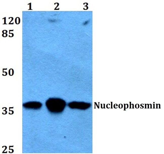
- Western blot analysis of Nucleophosmin using Nucleophosmin polyclonal antibody (Product # PA5-36279) at a dilution of 1:500. Lane 1: HEK293T whole cell lysate, Lane 2: Raw264.7 whole cell lysate, Lane 3: H9C2 whole cell lysate.
- Submitted by
- Invitrogen Antibodies (provider)
- Main image

- Experimental details
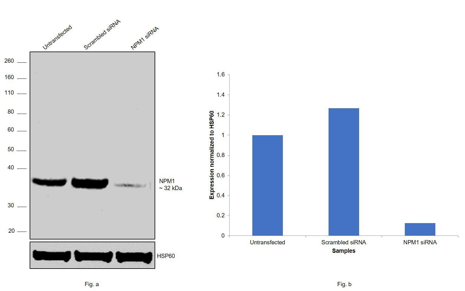
- Knockdown of NPM1 was achieved by transfecting HeLa with NPM1 specific siRNAs (Silencer® select Product # s9677, s9676). Western blot analysis (Fig. a) was performed using modified whole cell extracts (1% SDS) from NPM1 knockdown cells (lane 3), non-specific scrambled siRNA transfected cells (lane 2) and untransfected cells (lane 1). The blot was probed with NPM1 Polyclonal Antibody (Product # PA5-36279, 1:1200 dilution) and Goat anti-Rabbit IgG (H+L), Superclonal™ Recombinant Secondary Antibody, HRP (Product # A27036, 1:4000 dilution). Densitometric analysis of this western blot is shown in histogram (Fig. b). Decrease in signal upon siRNA mediated knock down confirms that antibody is specific to NPM1.
- Submitted by
- Invitrogen Antibodies (provider)
- Main image
- Experimental details
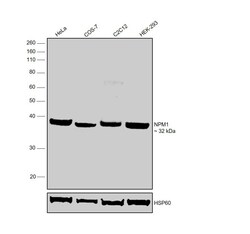
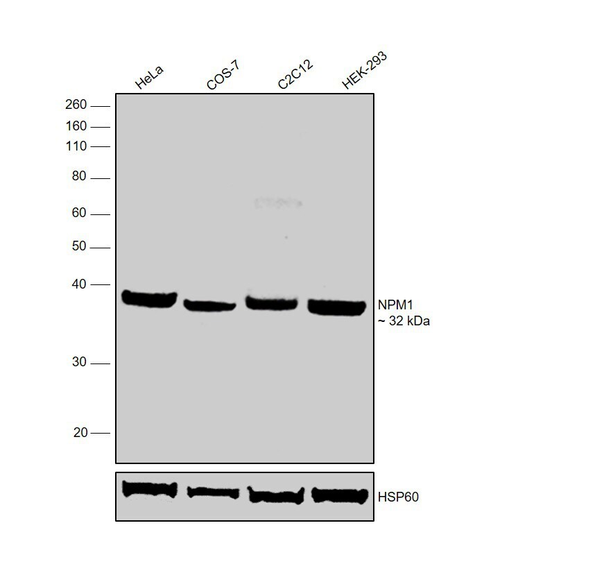
- Western blot was performed using Anti-NPM1 Polyclonal Antibody (Product # PA5-36279) and a 32 kDa band corresponding to NPM1 was observed across all the cell lines tested. Modified whole cell extracts (1% SDS) (30 µg lysate) of HeLa (Lane 1), COS-7 (Lane 2), C2C12 (Lane 3) and HEK-293 (Lane 4) were electrophoresed using NuPAGE™ 10% Bis-Tris Protein Gel (Product # NP0302BOX). Resolved proteins were then transferred onto a nitrocellulose membrane (Product # IB23001) by iBlot® 2 Dry Blotting System (Product # IB21001). The blot was probed with the primary antibody (1:1200 dilution) and detected by chemiluminescence with Goat anti-Rabbit IgG (H+L), Superclonal™ Recombinant Secondary Antibody, HRP (Product # A27036, 1:4000 dilution) using the iBright FL 1000 (Product # A32752). Chemiluminescent detection was performed using Novex® ECL Chemiluminescent Substrate Reagent Kit (Product # WP20005).
Supportive validation
- Submitted by
- Invitrogen Antibodies (provider)
- Main image
- Experimental details
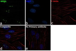
- Immunofluorescence analysis of NPM1 was performed using 70% confluent log phase HeLa cells. The cells were fixed with 4% Paraformaldehyde for 10 minutes, permeabilized with 0.1% Triton™ X-100 for 10 minutes, and blocked with 2% BSA for 10 minutes at room temperature. The cells were labeled with NPM1 Polyclonal Antibody (Product # PA5-36279) at 1:100 dilution in 0.1% BSA, incubated at 4 degree Celsius overnight and then labeled with Goat anti-Rabbit IgG (H+L) Superclonal™ Recombinant Secondary Antibody, Alexa Fluor® 488 conjugate (Product # A27034, 1:2000 dilution) for 45 minutes at room temperature (Panel a: Green). Nuclei (Panel b: Blue) were stained with ProLong™ Diamond Antifade Mountant with DAPI (Product # P36962). F-actin (Panel c: Red) was stained with Rhodamine Phalloidin (Product # R415, 1:300). Panel d represents the merged image showing nucleolar localization. Panel e represents control cells with no primary antibody to assess background. The images were captured at 60X magnification.

Supportive validation
- Submitted by
- Invitrogen Antibodies (provider)
- Main image
- Experimental details
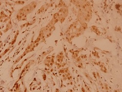
- Immunohistochemical analysis of Nucleophosmin in paraffin-embedded human breast carcinoma using Nucleophosmin polyclonal antibody (Product # PA5-36279) at a dilution of 1:100.

Supportive validation
- Submitted by
- Invitrogen Antibodies (provider)
- Main image
- Experimental details
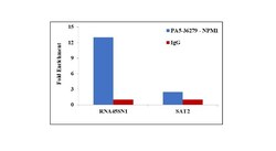
- Chromatin Immunoprecipitation (ChIP) assay of endogenous NPM1 protein using Anti-NPM1 Antibody: ChIP was performed using Anti-NPM1 Rabbit polyclonal Antibody (Product # PA5-36279, 5 µg) on sheared chromatin from HeLa cells using the MAGnify ChIP System kit (Product # 49-2024). Normal Rabbit IgG was used as a negative IP control. The purified DNA was analyzed by qPCR using primers binding to 45s rDNA gene body (+13kb) and SAT2 satellite repeats. Data is presented as fold enrichment of the antibody signal versus the negative control IgG using the comparative CT method.
