 Explore
 Validate
 Learn
Learn Western blot
 Immunocytochemistry
Antibody data
- Antibody Data
- Antigen structure
- References [4]
- Comments [0]
- Validations
- Immunocytochemistry [1]
Submit
Validation data
Reference
Comment
Report error
- Product number
- HPA011384 - Provider product page

- Provider
- Atlas Antibodies
- Proper citation
- Atlas Antibodies Cat#HPA011384, RRID:AB_1854692
- Product name
- Anti-NPM1
- Antibody type
- Polyclonal
- Description
- Polyclonal Antibody against Human NPM1, Gene description: nucleophosmin (nucleolar phosphoprotein B23, numatrin), Alternative Gene Names: B23, NPM, Validated applications: ICC, IHC, WB, Uniprot ID: P06748, Storage: Store at +4°C for short term storage. Long time storage is recommended at -20°C.
- Reactivity
- Human
- Host
- Rabbit
- Conjugate
- Unconjugated
- Isotype
- IgG
- Vial size
- 100 µl
- Concentration
- 0.1 mg/ml
- Storage
- Store at +4°C for short term storage. Long time storage is recommended at -20°C.
- Handling
- The antibody solution should be gently mixed before use.
Submitted references Host Factor Nucleophosmin 1 (NPM1/B23) Exerts Antiviral Effects against Chikungunya Virus by Its Interaction with Viral Nonstructural Protein 3
Roniciclib down-regulates stemness and inhibits cell growth by inducing nucleolar stress in neuroblastoma
The Proteomics of Colorectal Cancer: Identification of a Protein Signature Associated with Prognosis
Toponostics of invasive ductal breast carcinoma: combination of spatial protein expression imaging and quantitative proteome signature analysis.
Pradeep P, Sivakumar K, Sreekumar E, Pelka P
Microbiology Spectrum 2023;11(4)
Microbiology Spectrum 2023;11(4)
Roniciclib down-regulates stemness and inhibits cell growth by inducing nucleolar stress in neuroblastoma
Ognibene M, Pezzolo A
Scientific Reports 2020;10(1)
Scientific Reports 2020;10(1)
The Proteomics of Colorectal Cancer: Identification of a Protein Signature Associated with Prognosis
Addison C, O'Dwyer D, Ralton L, O'Shea A, Murray G
PLoS ONE 2011;6(11):e27718
PLoS ONE 2011;6(11):e27718
Toponostics of invasive ductal breast carcinoma: combination of spatial protein expression imaging and quantitative proteome signature analysis.
Röwer C, Ziems B, Radtke A, Schmitt O, Reimer T, Koy C, Thiesen HJ, Gerber B, Glocker MO
International journal of clinical and experimental pathology 2011 Mar 31;4(5):454-67
International journal of clinical and experimental pathology 2011 Mar 31;4(5):454-67
No comments: Submit comment
Supportive validation
- Submitted by
- Atlas Antibodies (provider)
- Main image
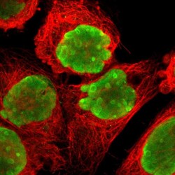
- Experimental details
- Immunofluorescent staining of human cell line A-431 shows localization to nucleus & nucleoli.
- Sample type
- Human
