Explore
 Validate
 Learn
Learn Western blot
Antibody data
- Antibody Data
- Antigen structure
- References [0]
- Comments [0]
- Validations
- Western blot [1]
- Immunocytochemistry [1]
- Immunohistochemistry [1]
- Flow cytometry [1]
Submit
Validation data
Reference
Comment
Report error
- Product number
- GTX22808 - Provider product page

- Provider
- GeneTex
- Proper citation
- GeneTex Cat#GTX22808, RRID:AB_384864
- Product name
- Spectrin beta 1 antibody [4C3]
- Antibody type
- Monoclonal
- Reactivity
- Human, Mouse, Rat
- Host
- Mouse
No comments: Submit comment
Supportive validation
- Submitted by
- GeneTex (provider)
- Main image
- Experimental details
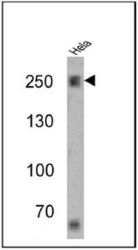
- Western blot analysis of Spectrin beta 1 in 25 ug of HeLa cell lysates. Proteins were transferred to a PVDF membrane and blocked at 4¢XC overnight. The membrane was probed with Spectrin beta 1 antibody [4C3] at a dilution of 1:1000 overnight at 4¢XC, washed in TBST, and probed with an HRP-conjugated secondary antibody for 1 hr at room temperature in the dark. Chemiluminescent detection was performed.

Supportive validation
- Submitted by
- GeneTex (provider)
- Main image
- Experimental details
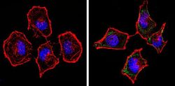
- Immunofluorescent analysis of Spectrin beta 1 (green) in HeLa cells (right) compared to a negative control without primary antibody (left). Formalin-fixed cells were permeabilized with 0.1% Triton X-100 in TBS for 5-10 minutes and blocked with 3% BSA-PBS for 30 minutes at room temperature. Cells were probed with Spectrin beta 1 antibody [4C3] in 3% BSA-PBS at a dilution of 1:100 and incubated overnight at 4 oC in a humidified chamber. Cells were washed with PBST and incubated with a proper secondary antibody in PBS at room temperature in the dark. F-actin (red) was stained with a flourescent red phalloidin and nuclei (blue) were stained with Hoechst or DAPI. Images were taken at a magnification of 60x.
Supportive validation
- Submitted by
- GeneTex (provider)
- Main image
- Experimental details
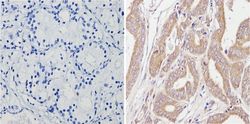
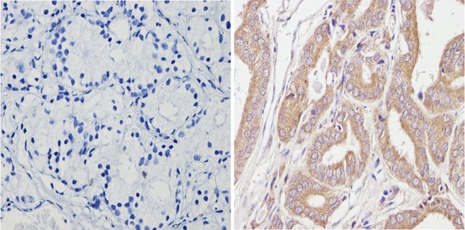
- Immunohistochemistry analysis of Spectrin beta 1 in paraffin-treated human prostate carcinoma (right) compared with a negative control in the absence of primary antibody (left). To expose target proteins, antigen retrieval method was performed using 10mM sodium citrate (pH 6.0) microwaved for 8-15 min. Following antigen retrieval, tissues were blocked in 3% H2O2-methanol for 15 min at room temperature, washed with ddH2O and PBS, and then probed with Spectrin beta 1 antibody [4C3] diluted by 3% BSA-PBS at a dilution of 1:20 overnight at 4¢XC in a humidified chamber. Tissues were washed extensively PBST and detection was performed using an HRP-conjugated secondary antibody followed by colorimetric detection using a DAB kit. Tissues were counterstained with hematoxylin and dehydrated with ethanol and xylene to prep for mounting.
Supportive validation
- Submitted by
- GeneTex (provider)
- Main image

- Experimental details
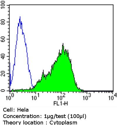
- Flow cytometry analysis of Spectrin beta 1 in HeLa cells compared to an isotype control (blue). Cells were harvested, adjusted to a concentration of 1-5x10^6 cells/ml, fixed with 2% paraformaldehyde and washed with PBS. Cells were penetrated by dropping the supernatant, adding 90% methanol and incubated for 10 minutes at room temperature. Follwing penetration, cells were blocked with a 2% solution of BSA-PBS for 30 min at room temperature and incubated with Spectrin beta 1 antibody [4C3] at a dilution of 1 ug/test for 60 min at room temperature. Cells were then incubated for 40 min at room temperature in the dark using a proper secondary antibody and re-suspended in PBS for FACS analysis.