Explore
 Validate
 Learn
Learn Western blot
 Immunocytochemistry
Antibody data
- Antibody Data
- Antigen structure
- References [0]
- Comments [0]
- Validations
- Immunocytochemistry [1]
- Chromatin Immunoprecipitation [1]
Submit
Validation data
Reference
Comment
Report error
- Product number
- 703926 - Provider product page

- Provider
- Invitrogen Antibodies
- Product name
- SMC3 Recombinant Rabbit Monoclonal Antibody (2H3L10)
- Antibody type
- Monoclonal
- Antigen
- Other
- Reactivity
- Human, Mouse
- Host
- Rabbit
- Isotype
- IgG
- Antibody clone number
- 2H3L10
- Vial size
- 100 µg
- Concentration
- 0.5 mg/mL
- Storage
- Store at 4°C short term. For long term storage, store at -20°C, avoiding freeze/thaw cycles.
No comments: Submit comment
Supportive validation
- Submitted by
- Invitrogen Antibodies (provider)
- Main image
- Experimental details
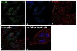
- For immunocytochemistry analysis, HeLa cells were fixed and permeabilized for detection of endogenous SMC3 using Anti-SMC3 Recombinant Rabbit Monoclonal Antibody (Product # 703926) at a 1:100 dilution and labeled with Goat anti-Rabbit IgG (H+L) Superclonal™ Secondary Antibody, Alexa Fluor® 488 conjugate (Product # A27034) at a 1:2000 dilution. Panel a) shows representative cells that were stained for detection and localization of SMC3 protein (green), Panel b) is stained for nuclei (blue) using ProLong™ Diamond Antifade Mountant with DAPI (Product # P36962). Panel c) represents cytoskeletal F-actin staining using Rhodamine Phalloidin (Product # R415) at a 1:300 dilution. Panel d) is a composite image of Panels a, b and c clearly demonstrating nuclear localization of SMC3. Panel e) represents control cells with no primary antibody to assess background. The images were captured at 60X magnification.

Supportive validation
- Submitted by
- Invitrogen Antibodies (provider)
- Main image
- Experimental details
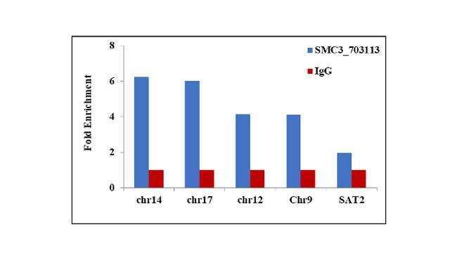
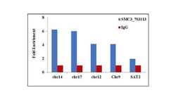
- Chromatin Immunoprecipitation (ChIP) assay of endogenous SMC3 protein using Anti-SMC3 Antibody: ChIP was performed using 5 µg of Anti-SMC3 Recombinant Rabbit Monoclonal Antibody (Product # 703926) on sheared chromatin from Hela cells using the MAGnify ChIP System kit (Product # 49-2024). Normal Rabbit IgG was used as a negative IP control. The purified DNA was analyzed by qPCR using primers binding to selected chromosomal positions: Chr 14 (75955167-75955445), Chr 17 (7944729-794507), Chr 12 (53746435-53746713), Chr 9 (11244 - 11320) and and SAT2 satellite repeats. Data is presented as fold enrichment of the antibody signal versus the negative control IgG using the comparative CT method.