Explore
 Validate
 Learn
Learn Western blot
Antibody data
- Antibody Data
- Antigen structure
- References [8]
- Comments [0]
- Validations
- Western blot [4]
- Immunoprecipitation [1]
- Immunohistochemistry [5]
Submit
Validation data
Reference
Comment
Report error
- Product number
- NB100-207 - Provider product page

- Provider
- Novus Biologicals
- Proper citation
- Novus Cat#NB100-207, RRID:AB_10002165
- Product name
- Rabbit Polyclonal SMC3 Antibody
- Antibody type
- Polyclonal
- Description
- Immunogen affinity purified.
- Reactivity
- Human, Mouse
- Host
- Rabbit
- Isotype
- IgG
- Vial size
- 100 ul
- Concentration
- 1.0 mg/ml
- Storage
- Store at 4C. Do not freeze.
Submitted references A Novel Role for α-Importins and Akirin in Establishment of Meiotic Sister Chromatid Cohesion in Caenorhabditis elegans.
HDAC8 mutations in Cornelia de Lange syndrome affect the cohesin acetylation cycle.
Two different replication factor C proteins, Ctf18 and RFC1, separately control PCNA-CRL4Cdt2-mediated Cdt1 proteolysis during S phase and following UV irradiation.
Human Timeless and Tipin stabilize replication forks and facilitate sister-chromatid cohesion.
Cohesin SMC1beta protects telomeres in meiocytes.
Cohesin acetylation speeds the replication fork.
Sororin, a substrate of the anaphase-promoting complex, is required for sister chromatid cohesion in vertebrates.
Cohesin SMC1 beta is required for meiotic chromosome dynamics, sister chromatid cohesion and DNA recombination.
Bowman R, Balukof N, Ford T, Smolikove S
Genetics 2019 Feb;211(2):617-635
Genetics 2019 Feb;211(2):617-635
HDAC8 mutations in Cornelia de Lange syndrome affect the cohesin acetylation cycle.
Deardorff MA, Bando M, Nakato R, Watrin E, Itoh T, Minamino M, Saitoh K, Komata M, Katou Y, Clark D, Cole KE, De Baere E, Decroos C, Di Donato N, Ernst S, Francey LJ, Gyftodimou Y, Hirashima K, Hullings M, Ishikawa Y, Jaulin C, Kaur M, Kiyono T, Lombardi PM, Magnaghi-Jaulin L, Mortier GR, Nozaki N, Petersen MB, Seimiya H, Siu VM, Suzuki Y, Takagaki K, Wilde JJ, Willems PJ, Prigent C, Gillessen-Kaesbach G, Christianson DW, Kaiser FJ, Jackson LG, Hirota T, Krantz ID, Shirahige K
Nature 2012 Sep 13;489(7415):313-7
Nature 2012 Sep 13;489(7415):313-7
Two different replication factor C proteins, Ctf18 and RFC1, separately control PCNA-CRL4Cdt2-mediated Cdt1 proteolysis during S phase and following UV irradiation.
Shiomi Y, Hayashi A, Ishii T, Shinmyozu K, Nakayama J, Sugasawa K, Nishitani H
Molecular and cellular biology 2012 Jun;32(12):2279-88
Molecular and cellular biology 2012 Jun;32(12):2279-88
Human Timeless and Tipin stabilize replication forks and facilitate sister-chromatid cohesion.
Leman AR, Noguchi C, Lee CY, Noguchi E
Journal of cell science 2010 Mar 1;123(Pt 5):660-70
Journal of cell science 2010 Mar 1;123(Pt 5):660-70
Cohesin SMC1beta protects telomeres in meiocytes.
Adelfalk C, Janschek J, Revenkova E, Blei C, Liebe B, Göb E, Alsheimer M, Benavente R, de Boer E, Novak I, Höög C, Scherthan H, Jessberger R
The Journal of cell biology 2009 Oct 19;187(2):185-99
The Journal of cell biology 2009 Oct 19;187(2):185-99
Cohesin acetylation speeds the replication fork.
Terret ME, Sherwood R, Rahman S, Qin J, Jallepalli PV
Nature 2009 Nov 12;462(7270):231-4
Nature 2009 Nov 12;462(7270):231-4
Sororin, a substrate of the anaphase-promoting complex, is required for sister chromatid cohesion in vertebrates.
Rankin S, Ayad NG, Kirschner MW
Molecular cell 2005 Apr 15;18(2):185-200
Molecular cell 2005 Apr 15;18(2):185-200
Cohesin SMC1 beta is required for meiotic chromosome dynamics, sister chromatid cohesion and DNA recombination.
Revenkova E, Eijpe M, Heyting C, Hodges CA, Hunt PA, Liebe B, Scherthan H, Jessberger R
Nature cell biology 2004 Jun;6(6):555-62
Nature cell biology 2004 Jun;6(6):555-62
No comments: Submit comment
Supportive validation
- Submitted by
- Novus Biologicals (provider)
- Main image

- Experimental details
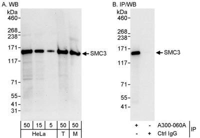
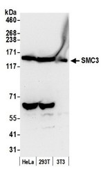
- Western Blot: SMC3 Antibody [NB100-207] - Samples: Whole cell lysate from HeLa (5, 15 and 50 ug for WB; 1 mg for IP, 20% of IP loaded), 293T (T; 50 ug) and mouse NIH3T3 (M; 50ug) cells. Affinity purified rabbit anti-SMC3 antibody used for WB at 0.1 ug/ml (A) and 1 ug/ml (B) and used for IP at 3 ug/mg lysate. Detection: Chemiluminescence with exposure times of 30 seconds (A) and 3 seconds (B).
- Submitted by
- Novus Biologicals (provider)
- Main image
- Experimental details
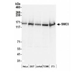
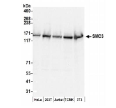
- Western Blot: SMC3 Antibody [NB100-207] - Whole cell lysate (50 ug) from HeLa, 293T, Jurkat, mouse TCMK-1, and mouse NIH3T3 cells prepared using NETN lysis buffer. Antibodies: Affinity purified rabbit anti-SMC3 antibody used for WB at 0.1 ug/ml. Detection: Chemiluminescence with an exposure time of 3 seconds.
- Submitted by
- Novus Biologicals (provider)
- Main image
- Experimental details
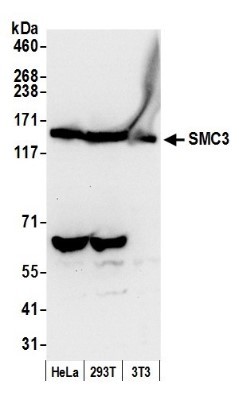
- Western Blot: SMC3 Antibody [NB100-207] - Samples: Whole cell lysate (50 ug) from HeLa, 293T, and mouse NIH3T3 cells prepared using NETN lysis buffer. Antibody: Affinity purified rabbit anti-SMC3 antibody used for WB at 0.1 ug/ml. Detection: Chemiluminescence with an exposure time of 10 seconds.
- Submitted by
- Novus Biologicals (provider)
- Main image
- Experimental details
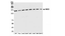
- Western Blot: SMC3 Antibody [NB100-207] - Whole cell lysate (10 ug) from HEK293T, HeLa, Jurkat, LNCaP, U2OS, A-549, TCMK-1, and NIH 3T3 cells prepared using NETN lysis buffer. Antibody: Affinity purified rabbit anti-SMC3 antibody used for WB at 0.04 ug/ml. Detection: Chemiluminescence with an exposure time of 3 seconds.

Supportive validation
- Submitted by
- Novus Biologicals (provider)
- Main image
- Experimental details
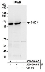
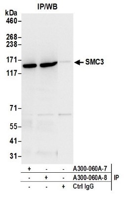
- Immunoprecipitation: SMC3 Antibody [NB100-207] - Samples: Whole cell lysate (0.5 or 1.0 mg per IP reaction; 20% of IP loaded) from HeLa cells prepared using NETN lysis buffer. Antibodies: Affinity purified rabbit anti-SMC3 antibody (lot was used at 1 ug/ml. Detection: Chemiluminescence with an exposure time of 3 seconds.
Supportive validation
- Submitted by
- Novus Biologicals (provider)
- Main image
- Experimental details
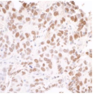
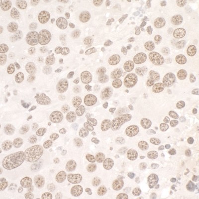
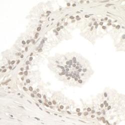
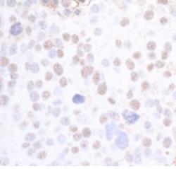
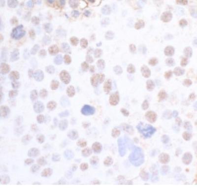
- Immunohistochemistry-Paraffin: SMC3 Antibody [NB100-207] - FFPE section of human testicular seminoma. Antibody: Affinity purified rabbit anti-SMC3 used at a dilution of 1:1,000 (1ug/ml). Detection: DAB
- Submitted by
- Novus Biologicals (provider)
- Main image

- Experimental details
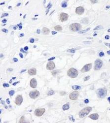
- Immunohistochemistry: SMC3 Antibody [NB100-207] - Sample: FFPE section of mouse renal cell carcinoma. Antibody: Affinity purified rabbit anti-SMC3 Cat. No. Lot8 used at a dilution of 1:1,000 (1ug/ml). Detection: DAB. Counterstain: IHC Hematoxylin (blue).
- Submitted by
- Novus Biologicals (provider)
- Main image
- Experimental details
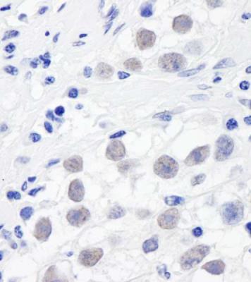
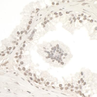
- Immunohistochemistry: SMC3 Antibody [NB100-207] - Sample: FFPE section of human prostate carcinoma. Antibodies: Affinity purified rabbit anti-SMC3 Cat. No. Lot8 used at a dilution of 1:5,000 (0.2ug/ml). Detection: DAB. Counterstain: IHC Hematoxylin (blue).
- Submitted by
- Novus Biologicals (provider)
- Main image
- Experimental details
- Immunohistochemistry-Paraffin: SMC3 Antibody [NB100-207] - Section of mouse renal cell carcinoma. Antibody: Affinity purified rabbit anti-SMC3 used at 1:5,000 (0.2ug/ml). Detection: DAB
- Submitted by
- Novus Biologicals (provider)
- Main image

- Experimental details
- Immunohistochemistry-Paraffin: SMC3 Antibody [NB100-207] - Section of human breast carcinoma. Antibody: Affinity purified rabbit anti-SMC3 used at 1:5,000 (0.2ug/ml). Detection: DAB