Explore
 Validate
 Learn
Learn Western blot
 Immunocytochemistry
Antibody data
- Antibody Data
- Antigen structure
- References [0]
- Comments [0]
- Validations
- Immunocytochemistry [4]
- Immunohistochemistry [4]
- Other assay [1]
Submit
Validation data
Reference
Comment
Report error
- Product number
- PA5-52629 - Provider product page

- Provider
- Invitrogen Antibodies
- Product name
- HEXIM1 Polyclonal Antibody
- Antibody type
- Polyclonal
- Antigen
- Recombinant protein fragment
- Description
- Immunogen sequence: AKSDDTSDDD FMEEGGEEDG GSDGMGGDGS EFLQRDFSET YERYHTESLQ NMSKQELIKE YLELEKCLSR MEDENNRLRL ESKRLGGDDA RVRELELELD RLRAENLQLL TENELHRQQE RA Highest antigen sequence identity to the following orthologs: Mouse - 95%, Rat - 94%.
- Reactivity
- Human
- Host
- Rabbit
- Isotype
- IgG
- Vial size
- 100 μL
- Concentration
- 0.4 mg/mL
- Storage
- Store at 4°C short term. For long term storage, store at -20°C, avoiding freeze/thaw cycles.
No comments: Submit comment
Supportive validation
- Submitted by
- Invitrogen Antibodies (provider)
- Main image
- Experimental details
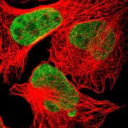
- Immunofluorescent staining of HEXIM1 in human cell line U-2 OS shows positivity in nucleus but excluded from the nucleoli. Samples were probed using a HEXIM1 Polyclonal Antibody (Product # PA5-52629).

- Submitted by
- Invitrogen Antibodies (provider)
- Main image
- Experimental details
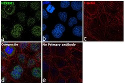
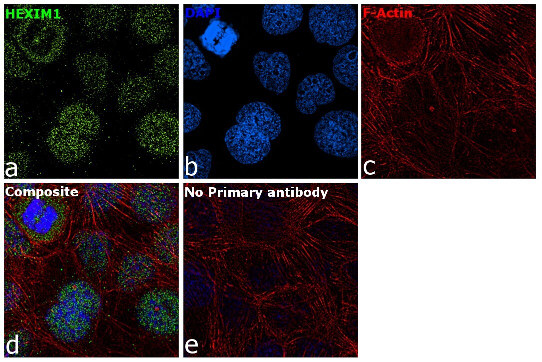
- Immunofluorescence analysis of HEXIM1 was performed using A-431 cells. The cells were fixed with 4% paraformaldehyde for 10 minutes, permeabilized with 0.1% Triton™ X-100 for 15 minutes, and blocked with 1% BSA for 1 hour at room temperature. The cells were labeled with HEXIM1 Polyclonal Antibody (Product # PA5-52629) at 2 µg/mL in 0.1% BSA, incubated at 4 degree Celsius overnight and then labeled with Goat anti-Rabbit IgG (H+L) Superclonal™ Secondary Antibody, Alexa Fluor® 488 conjugate (Product # A27034) at a dilution of 1:2000 for 45 minutes at room temperature (Panel a: green). Nuclei (Panel b: blue) were stained with ProLong™ Diamond Antifade Mountant with DAPI (Product # P36962). F-actin (Panel c: red) was stained with Rhodamine Phalloidin (Product # R415, 1:300). Panel d represents the merged image showing nuclear localization. Panel e represents control cells with no primary antibody to assess background. The images were captured at 60X magnification..

- Submitted by
- Invitrogen Antibodies (provider)
- Main image
- Experimental details
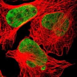
- Immunofluorecent analysis of HEXIM1 in human cell line U-2 OS using HEXIM1 Polyclonal Antibody (Product # PA5-52629). Staining shows localization to nucleoplasm.

- Submitted by
- Invitrogen Antibodies (provider)
- Main image

- Experimental details
- Immunofluorescence analysis of HEXIM1 was performed using A-431 cells. The cells were fixed with 4% paraformaldehyde for 10 minutes, permeabilized with 0.1% Triton™ X-100 for 15 minutes, and blocked with 1% BSA for 1 hour at room temperature. The cells were labeled with HEXIM1 Polyclonal Antibody (Product # PA5-52629) at 2 µg/mL in 0.1% BSA, incubated at 4 degree Celsius overnight and then labeled with Goat anti-Rabbit IgG (Heavy Chain) Superclonal™ Secondary Antibody, Alexa Fluor® 488 conjugate (Product # A27034) at a dilution of 1:2000 for 45 minutes at room temperature (Panel a: green). Nuclei (Panel b: blue) were stained with ProLong™ Diamond Antifade Mountant with DAPI (Product # P36962). F-actin (Panel c: red) was stained with Rhodamine Phalloidin (Product # R415, 1:300). Panel d represents the merged image showing nuclear localization. Panel e represents control cells with no primary antibody to assess background. The images were captured at 60X magnification..
Supportive validation
- Submitted by
- Invitrogen Antibodies (provider)
- Main image
- Experimental details
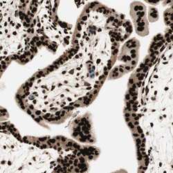
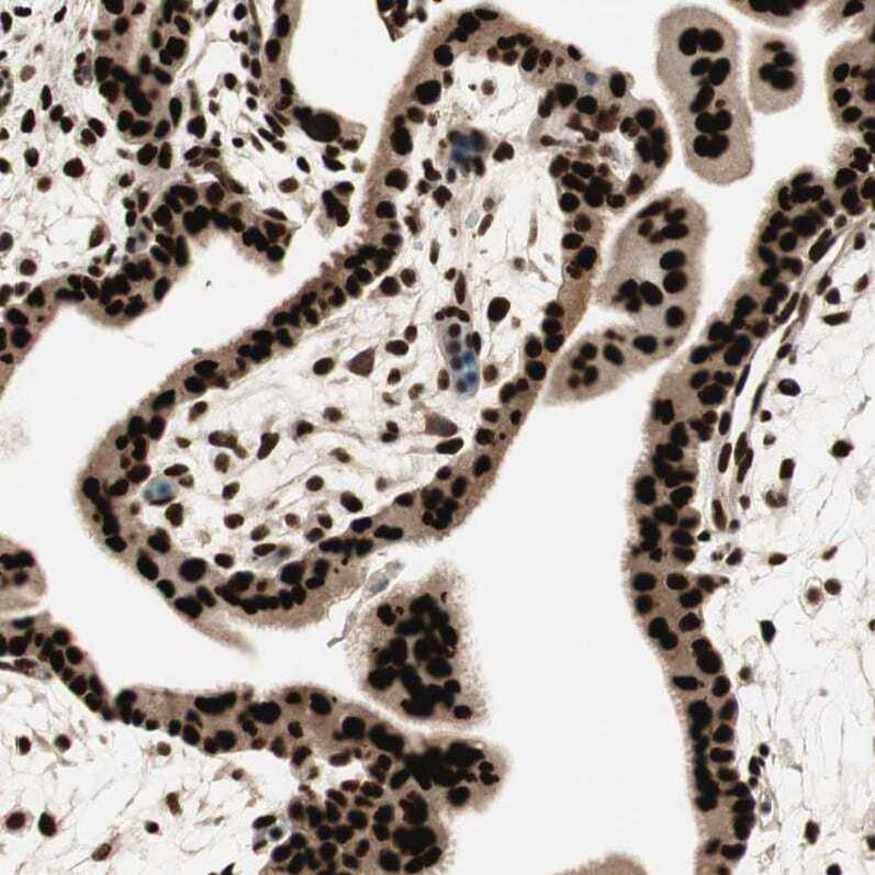
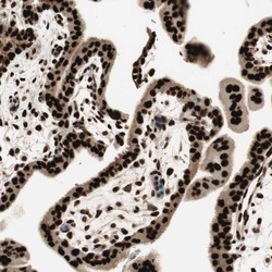
- Immunohistochemical analysis of HEXIM1 in human placenta using HEXIM1 Polyclonal Antibody (Product # PA5-52629) shows strong nuclear positivity in trophoblastic cells.
- Submitted by
- Invitrogen Antibodies (provider)
- Main image
- Experimental details
- Immunohistochemical staining of HEXIM1 in human placenta using a HEXIM1 Polyclonal Antibody (Product # PA5-52629) shows strong nuclear positivity in trophoblastic cells.
- Submitted by
- Invitrogen Antibodies (provider)
- Main image
- Experimental details
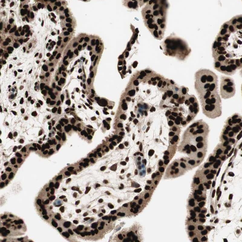
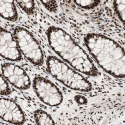
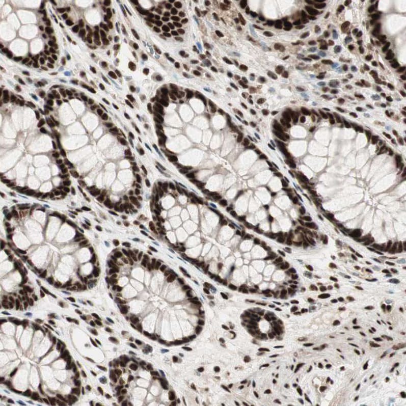
- Immunohistochemical analysis of HEXIM1 in human colon using HEXIM1 Polyclonal Antibody (Product # PA5-52629) shows strong nuclear positivity in glandular cells.
- Submitted by
- Invitrogen Antibodies (provider)
- Main image
- Experimental details
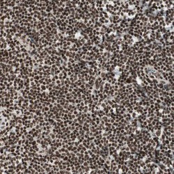
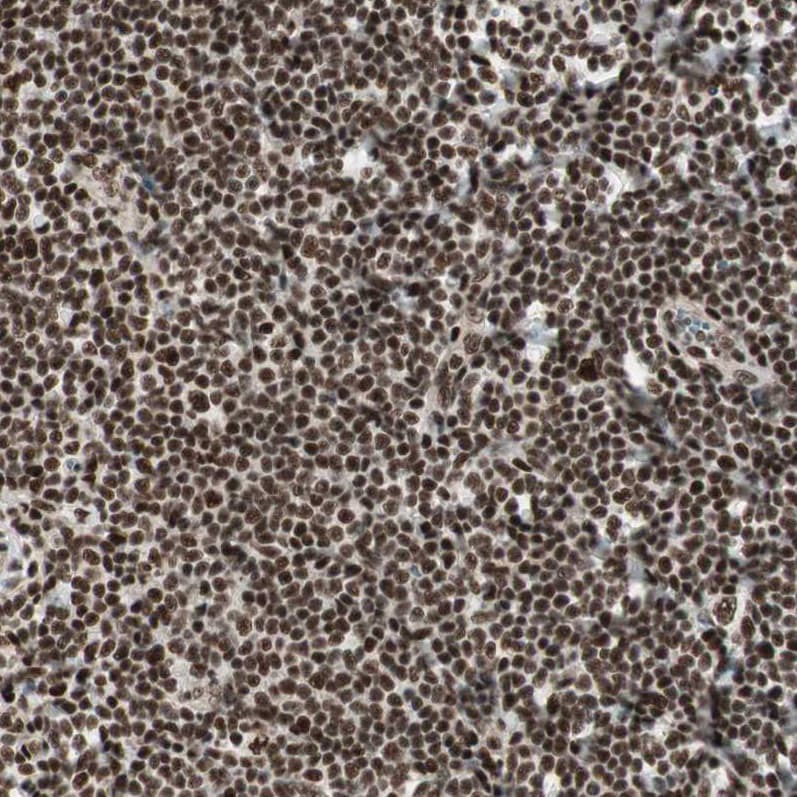
- Immunohistochemical analysis of HEXIM1 in human tonsil using HEXIM1 Polyclonal Antibody (Product # PA5-52629) shows strong nuclear positivity in non-germinal center cells.
Supportive validation
- Submitted by
- Invitrogen Antibodies (provider)
- Main image

- Experimental details
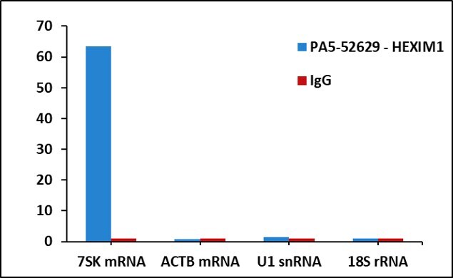
- Detection of binding of endogenous HEXIM1 protein to specific RNA using Anti-HEXIM1 Antibody: RNA Immunoprecipitation (RIP) was performed using Anti-HEXIM1 Recombinant Rabbit Polyclonal Antibody (Product # PA5-52629, 5 µg) on whole cell lysate from 2 million HCT 116 cells. Normal Rabbit IgG was used as a negative IP control. RNA purified by RiboPure™ RNA Purification Kit (Product # AM1924) was analyzed by RT-PCR using the Power SYBR® Green RNA-to-CT™ 1-Step Kit (Product # 4389986) with RIP primer pairs over 7SK, ACTB mRNA, U1 snRNA and 18s rRNA. Data is presented as fold enrichment of the antibody signal versus the negative control IgG using the comparative CT method.