 Explore
 Validate
 Learn
Learn Western blot
Antibody data
- Antibody Data
- Antigen structure
- References [0]
- Comments [0]
- Validations
- Western blot [4]
- Immunocytochemistry [2]
- Immunohistochemistry [5]
Submit
Validation data
Reference
Comment
Report error
- Product number
- PA5-53251 - Provider product page

- Provider
- Invitrogen Antibodies
- Product name
- SSR3 Polyclonal Antibody
- Antibody type
- Polyclonal
- Antigen
- Recombinant full-length protein
- Description
- Immunogen sequence: VLKHKVAQKR EDAVSKEVTR KLSEADNRKM SRKEKDERIL WKKNEVADYE A
- Concentration
- 0.1 mg/mL
No comments: Submit comment
Supportive validation
- Submitted by
- Invitrogen Antibodies (provider)
- Main image

- Experimental details
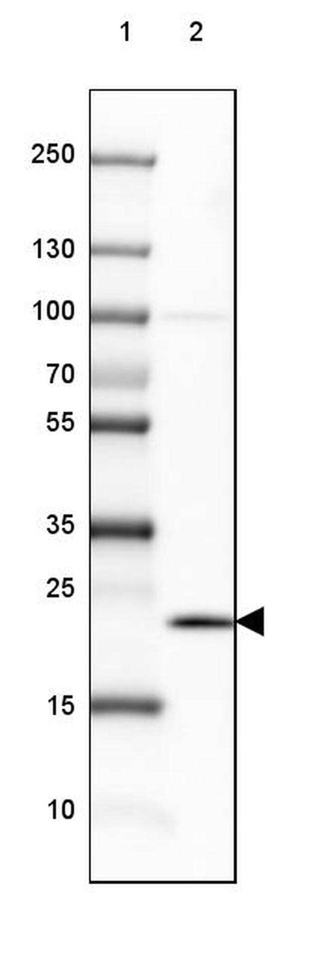
- Western blot analysis of SSR3 in Lane 1: Marker (kDa) 250, 130, 100, 70, 55, 35, 25, 15, 10; Lane 2: Human cerebral cortex tissue. Samples were probed using a SSR3 Polyclonal Antibody (Product # PA5-53251).
- Submitted by
- Invitrogen Antibodies (provider)
- Main image
- Experimental details
- Western blot analysis of SSR3 in Lane 1: Marker (kDa) 250, 130, 100, 70, 55, 35, 25, 15, 10; Lane 2: Mouse cerebellum tissue. Samples were probed using a SSR3 Polyclonal Antibody (Product # PA5-53251).

- Submitted by
- Invitrogen Antibodies (provider)
- Main image
- Experimental details
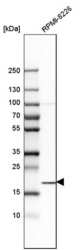
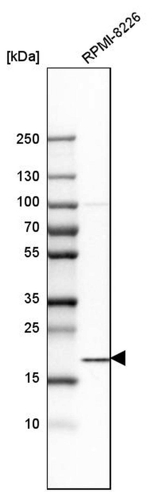
- Western blot analysis of SSR3 in human cell line RPMI-8226 using a SSR3 Polyclonal Antibody (Product # PA5-53251).
- Submitted by
- Invitrogen Antibodies (provider)
- Main image
- Experimental details
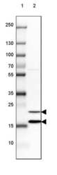
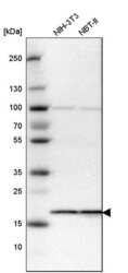
- Western blot analysis of SSR3 in mouse cell line NIH-3T3 and rat cell line NBT-II using a SSR3 Polyclonal Antibody (Product # PA5-53251).

Supportive validation
- Submitted by
- Invitrogen Antibodies (provider)
- Main image

- Experimental details
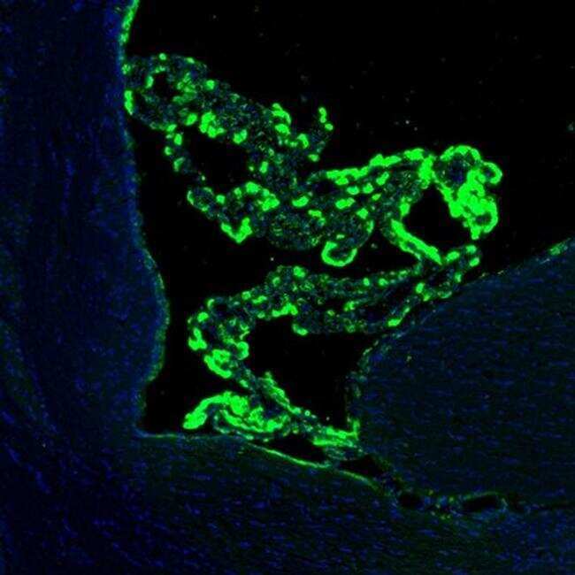
- Immunofluorescent staining of SSR3 in mouse choroid plexus shows strong immunoreactivity in a subset of cells. Samples were probed using a SSR3 Polyclonal Antibody (Product # PA5-53251).
- Submitted by
- Invitrogen Antibodies (provider)
- Main image
- Experimental details
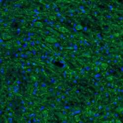
- Immunofluorescent staining of SSR3 in mouse trigeminal nucleus shows positivity in neurons and their processes. Samples were probed using a SSR3 Polyclonal Antibody (Product # PA5-53251).
Supportive validation
- Submitted by
- Invitrogen Antibodies (provider)
- Main image

- Experimental details
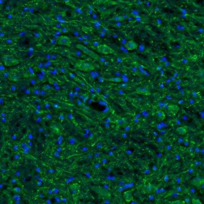
- Immunofluorescent staining of SSR3 in mousebasal forebrain shows weak labeling in the caudate putamen. Samples were probed using a SSR3 Polyclonal Antibody (Product # PA5-53251).
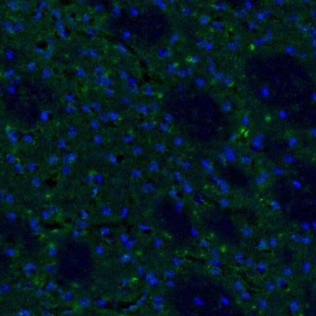
- Submitted by
- Invitrogen Antibodies (provider)
- Main image

- Experimental details
- Immunofluorescent staining of SSR3 in mouse brain shows strong immunoreactivity in median eminens. Samples were probed using a SSR3 Polyclonal Antibody (Product # PA5-53251).

- Submitted by
- Invitrogen Antibodies (provider)
- Main image
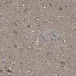
- Experimental details
- Immunohistochemical staining of SSR3 in human lateral ventricle shows weak positivity in neuropil and a few neurons. Samples were probed using a SSR3 Polyclonal Antibody (Product # PA5-53251).

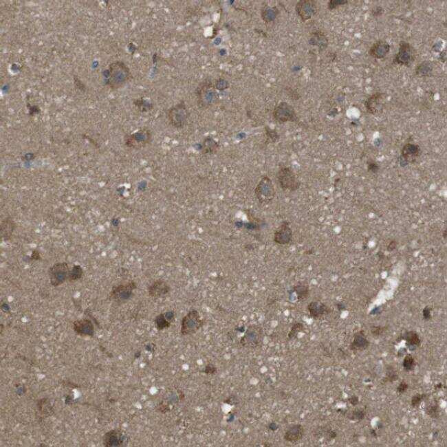
- Submitted by
- Invitrogen Antibodies (provider)
- Main image

- Experimental details
- Immunohistochemical staining of SSR3 in human cerebral cortex tissue shows moderate cytoplasmic positivity in neurons. Samples were probed using a SSR3 Polyclonal Antibody (Product # PA5-53251).
- Submitted by
- Invitrogen Antibodies (provider)
- Main image
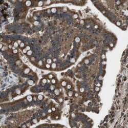
- Experimental details
- Immunohistochemical staining of SSR3 in human colon tissue shows strong cytoplasmic positivity in glandular cells. Samples were probed using a SSR3 Polyclonal Antibody (Product # PA5-53251).
