Explore
 Validate
 Learn
Learn Western blot
 ELISA
ELISAAntibody data
- Antibody Data
- Antigen structure
- References [0]
- Comments [0]
- Validations
- Western blot [1]
- Immunohistochemistry [1]
Submit
Validation data
Reference
Comment
Report error
- Product number
- AP09130PU-N - Provider product page

- Provider
- Acris Antibodies GmbH
- Proper citation
- Acris Antibodies GmbH Cat#AP09130PU-N, RRID:AB_2035391
- Product name
- anti Cyclin L1 (Isoform 1)
- Antibody type
- Polyclonal
- Antigen
- Synthetic polypeptide corresponding to amino acids 314-369 of Human Cyclin L1alpha protein
- Reactivity
- Human
- Host
- Rabbit
- Isotype
- IgG
- Vial size
- 0.5 mg
- Concentration
- 5.0 mg/ml (by UV absorbance at 280 nm)
No comments: Submit comment
Supportive validation
- Submitted by
- Acris Antibodies GmbH (provider)
- Main image

- Experimental details
- Western blot using Protein A Purified anti-Cyclin L1alpha antibody shows detection of a band ~59 kDa corresponding to a Cyclin L1alpha (arrowhead) present in mouse brain whole cell lysate. Approximately 35 µg of lysate was separated by 4-20% SDS-PAGE followed by transfer to nitrocellulose. After blocking the membrane was probed with the primary antibody diluted to 1:3,500 for 2h at room temperature followed by washes and reaction with a 1:10,000 dilution of IRDye(TM)800 conjugated Gt-a-Rabbit IgG [H&L] MX for 45 min at room temperature. IRDye(TM)800 fluorescence image was captured using the Odyssey(R) Infrared Imaging System developed by LI-COR. IRDye is a trademark of LI-COR, Inc. Other detection systems will yield similar results.

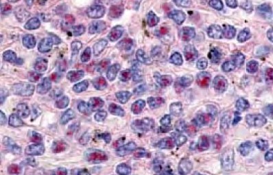
Supportive validation
- Submitted by
- Acris Antibodies GmbH (provider)
- Main image
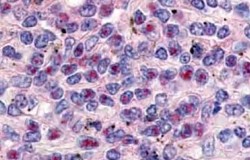
- Experimental details
- Immunohistochemistry. Anti-Cyclin L1alpha antibody was used at a 10 µg/ml to detect Cyclin L1alpha in a variety of tissues including breast (collagen), heart, kidney (distal tubules), liver, skeletal muscle, ovary (granulosa and oocyte), pancreas (islet), placenta (trophoblast), prostate (epithelium), skin, spleen (endothelium), stomach (chief), testes (seminiferous epithelium and leydig), thymus (Hassals corpuscle and lymphocytes) and uterus (glandular epithelium and stroma). Low to moderate background staining was noted. This image shows Cyclin L1alpha staining of human spleen. Tissue was formalin-fixed and paraffin embedded.