 Explore
 Validate
 Learn
Learn Western blot
Antibody data
- Antibody Data
- Antigen structure
- References [0]
- Comments [0]
- Validations
- Western blot [7]
- Immunocytochemistry [2]
- Chromatin Immunoprecipitation [1]
- Other assay [1]
Submit
Validation data
Reference
Comment
Report error
- Product number
- MA1-25451 - Provider product page

- Provider
- Invitrogen Antibodies
- Product name
- HDAC2 Monoclonal Antibody (HDAC2-62)
- Antibody type
- Monoclonal
- Antigen
- Synthetic peptide
- Description
- Recommended positive controls: NIH3T3.
- Antibody clone number
- HDAC2-62
- Concentration
- 2.5 mg/mL
No comments: Submit comment
Supportive validation
- Submitted by
- Invitrogen Antibodies (provider)
- Main image

- Experimental details
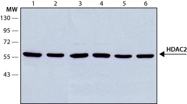
- Western blot analysis of HDAC2 in (lane 1) HEK-293T (lane 2) HeLa (lane 3) Jurkat (lane 4) K562 (lane 5) Neuro-2a (lane 6) NIH-3T3 lysates. Samples were separated on SDS-PAGE and probed withHDAC2 monoclonal antibody (Product # MA1-25451) at a dilution of 0.5 µg/mL. The antibody was developed using a Goat Anti-mouse IgG-Peroxidase and a chemiluminescent substrate.
- Submitted by
- Invitrogen Antibodies (provider)
- Main image
- Experimental details
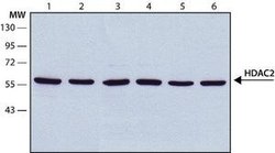
- Western Blot analysis of HDAC2 was performed by loading (1)HEK-293T (2) HeLa (3) JURKAT (4) K562 (5) Neuro-2a (6) NIH-3T3 cell lysates. Proteins were transferred to a membrane and probed with a HDAC2 Monoclonal Antibody (HDAC2-62) (Product # MA1-25451) at a dilution of 0.5 µg/mL.
- Submitted by
- Invitrogen Antibodies (provider)
- Main image
- Experimental details
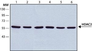
- Western Blot analysis of HDAC2 was performed by loading (1)HEK-293T (2) HeLa (3) JURKAT (4) K562 (5) Neuro-2a (6) NIH-3T3 cell lysates. Proteins were transferred to a membrane and probed with a HDAC2 Monoclonal Antibody (HDAC2-62) (Product # MA1-25451) at a dilution of 0.5 µg/mL.

- Submitted by
- Invitrogen Antibodies (provider)
- Main image

- Experimental details
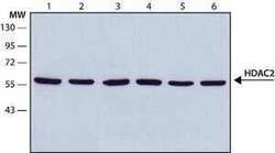
- Western Blot analysis of HDAC2 was performed by loading (1)HEK-293T (2) HeLa (3) JURKAT (4) K562 (5) Neuro-2a (6) NIH-3T3 cell lysates. Proteins were transferred to a membrane and probed with a HDAC2 Monoclonal Antibody (HDAC2-62) (Product # MA1-25451) at a dilution of 0.5 µg/mL.

- Submitted by
- Invitrogen Antibodies (provider)
- Main image
- Experimental details
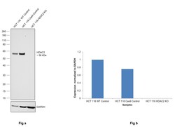
- Knockout of HDAC2 was achieved by CRISPR-Cas9 genome editing using LentiArray™ Lentiviral sgRNA (Product # A32042, Assay ID CRISPR1097024_LV) and LentiArray Cas9 Lentivirus (Product # A32064). Western blot analysis of HDAC2 was performed by loading 30 µg of HCT 116 wild type (Lane 1), HCT 116 Cas9 (Lane 2) andHCT 116 HDAC2 KO (Lane 3) whole cell extracts. The samples were electrophoresed using NuPAGE™ Novex™ 4-12% Bis-Tris Protein Gel (Product # NP0321BOX). Resolved proteins were then transferred onto a nitrocellulose membrane (Product # IB23001) by iBlot® 2 Dry Blotting System (Product # IB21001). The blot was probed with Anti-HDAC2 Monoclonal Antibody (HDAC2-62) (Product # MA1-25451, 0.5 µg/mL dilution) and Goat anti-Mouse IgG (H+L) Superclonal™ Recombinant Secondary Antibody, HRP (Product # A28177, 1:5000 dilution) using the iBright™ FL 1500 (Product # A44115). Chemiluminescent detection was performed using Novex® ECL Chemiluminescent Substrate Reagent Kit (Product # WP20005). Loss of signal upon CRISPR mediated knockout (KO) using the LentiArray™ CRISPR product line confirms that antibody is specific to HDAC2.
- Submitted by
- Invitrogen Antibodies (provider)
- Main image
- Experimental details
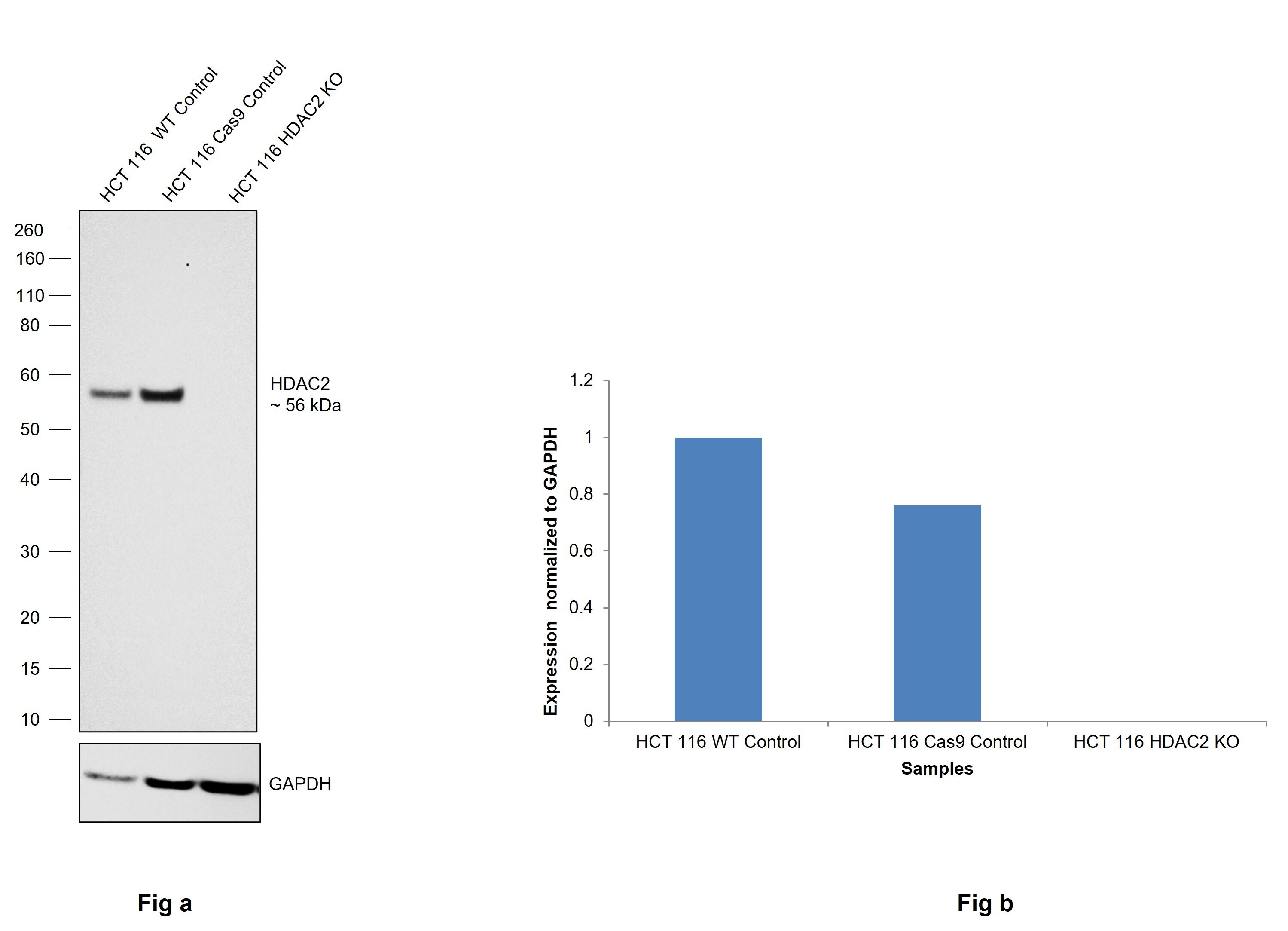
- Knockdown of HDAC2 was achieved by transfecting HEK-293 cells with HDAC2 specific siRNAs (Silencer® select Product # s6494). Western blot analysis (Fig. a) was performed using whole cell extracts from the HDAC2 knockdown cells (Lane 3), non-specific scrambled siRNA transfected cells (Lane 2) and untransfected cells (Lane 1). The blots were probed with Anti-HDAC2 Monoclonal Antibody (HDAC2-62) (Product # MA1-25451, 0.5 ug/ml) and Goat anti-Mouse IgG (H+L), Superclonal™ Recombinant Secondary Antibody, HRP (Product # A28177, 1:4000 dilution) using the iBright FL 1000 (Product # A32752). Densitometric analysis of this Western Blot is shown in histogram (Fig. b). Decrease in signal upon siRNA mediated knock down confirms that antibody is specific to HDAC2.

- Submitted by
- Invitrogen Antibodies (provider)
- Main image

- Experimental details
- Western blot was performed using Anti-HDAC2 Monoclonal Antibody (HDAC2-62) (Product # MA1-25451) and a 56 kDa band corresponding to HDAC2 was observed across the cell lines except MCF 10A which is reported to be negative. (Fig. a) Modified whole cell extracts (1% SDS) (30 µg lysate) of DU 145 (Lane 1), A549 (Lane 2), PC-3 (Lane 3), Hep G2 (Lane 4), HeLa (Lane 5), HEK-293 (Lane 6), K-562 (Lane 7), SH-SY5Y (Lane 8) and tissue extract (30 µg lysate) of Mouse Thymus; (Fig. b) Modified whole cell extracts (1% SDS) (30 µg lysate) of MCF7 (Lane 1), MDA-MB-231 (Lane 2), SK-BR-3 (Lane 3), T-47D (Lane 4) and MCF 10A were electrophoresed using Novex® NuPAGE® 4-12 % Bis-Tris gel (Product # NP0321BOX). Resolved proteins were then transferred onto a nitrocellulose membrane (Product # IB23001) by iBlot® 2 Dry Blotting System (Product # IB21001). The blot was probed with the primary antibody (0.5 ug/ml) and detected by chemiluminescence with Goat anti-Mouse IgG (H+L), Superclonal™ Recombinant Secondary Antibody, HRP (Product # A28177, 1:4000 dilution) using the iBright FL 1000 (Product # A32752). Chemiluminescent detection was performed using Novex® ECL Chemiluminescent Substrate Reagent Kit (Product # WP20005).

Supportive validation
- Submitted by
- Invitrogen Antibodies (provider)
- Main image
- Experimental details
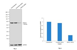
- Immunocytochemistry-Immunofluorescence analysis of HDAC2 in HeLa cells using HDAC2 Monoclonal Antibody (HDAC2-62) (Product # MA1-25451) at 10 µg/mL. Cells were fixed and permeabilized with cold methanol followed by cold methanol: acetone.

- Submitted by
- Invitrogen Antibodies (provider)
- Main image

- Experimental details
- Immunofluorescence analysis of HDAC2 was performed using 70% confluent log phase HeLa cells. The cells were fixed with 4% paraformaldehyde for 10 minutes, permeabilized with 0.1% Triton™ X-100 for 15 minutes, and blocked with 2% BSA for 1 hour at room temperature. The cells were labeled with HDAC2 Monoclonal Antibody (HDAC2-62) (Product MA1-25451) at 2 µg/mL in 0.1% BSA, incubated at 4 degree Celsius overnight and then with Goat anti-Mouse IgG (H+L), Superclonal™ Recombinant Secondary Antibody, Alexa Fluor 488 (Product # A28175) at a dilution of 1:2000 for 45 minutes at room temperature (Panel a: Green). Nuclei (Panel b: Blue) were stained with SlowFade® Gold Antifade Mountant with DAPI (Product # S36938). F-actin (Panel c: Red) was stained with Rhodamine Phalloidin (Product # R415, 1:300). Panel d represents the merged image showing nuclear localization. Panel e represents control cells with no primary antibody to assess background. The images were captured at 60X magnification.

Supportive validation
- Submitted by
- Invitrogen Antibodies (provider)
- Main image
- Experimental details
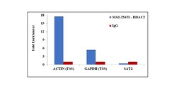
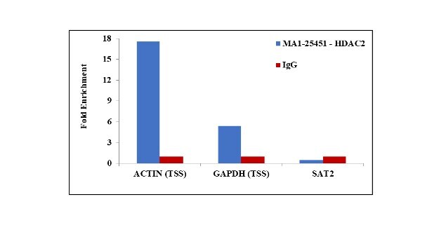
- Chromatin Immunoprecipitation (ChIP) assay of endogenous HDAC2 protein using Anti-HDAC2 Antibody: ChIP was performed using Anti-HDAC2 Mouse Monoclonal Antibody (Product # MA1-25451, 2.5 µg) on sheared chromatin from HeLa cells using the MAGnify ChIP System kit (Product # 49-2024). Normal Mouse IgG was used as a negative IP control. The purified DNA was analyzed by qPCR using primers binding to ACTIN and GAPDH transcriptional start site and SAT2 satellite repeats. Data is presented as fold enrichment of the antibody signal versus the negative control IgG using the comparative CT method.
Supportive validation
- Submitted by
- Invitrogen Antibodies (provider)
- Main image
- Experimental details
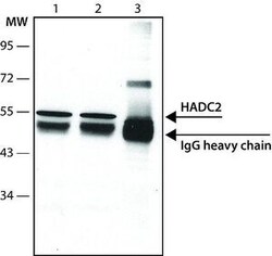
- Immunoprecipitation analysis of HDAC2 in HeLa cell lysates using a HDAC2 Monoclonal Antibody (HDAC2-62) (Product # MA1-25451). Lane 1 : IP from HeLa cell lysate using 5 µg antibody, lane 2 : IP from HeLa cell lysate using 2.5 µg antibody, lane 3 : IP from HeLa cell lysate using 5 µg non-relevant antibody.
