 Explore
 Validate
 Learn
LearnMA5-18061
antibody from Invitrogen Antibodies
Targeting: HDAC2
KDAC2, RPD3, YAF1
 Western blot Immunocytochemistry
 Immunoprecipitation Immunohistochemistry Flow cytometry Chromatin Immunoprecipitation
Antibody data
- Antibody Data
- Antigen structure
- References [0]
- Comments [0]
- Validations
- Western blot [4]
- Immunocytochemistry [1]
- Chromatin Immunoprecipitation [1]
Submit
Validation data
Reference
Comment
Report error
- Product number
- MA5-18061 - Provider product page

- Provider
- Invitrogen Antibodies
- Product name
- HDAC2 Monoclonal Antibody (3F3)
- Antibody type
- Monoclonal
- Antigen
- Synthetic peptide
- Description
- Western blot results show a band at 55 kDa, which matches the predicted molecular weight. This antibody is predicted to detect HDAC2 in Chicken and Bovine samples.
- Reactivity
- Human, Mouse, Rat
- Host
- Mouse
- Isotype
- IgG
- Antibody clone number
- 3F3
- Vial size
- 100 µL
- Concentration
- 1 mg/mL
- Storage
- Store at 4°C short term. For long term storage, store at -20°C, avoiding freeze/thaw cycles.
No comments: Submit comment
Supportive validation
- Submitted by
- Invitrogen Antibodies (provider)
- Main image

- Experimental details
- Western blot analysis of HDAC2 in Hela lysates using an HDAC2 monoclonal antibody (Product # MA5-18061) at various dilutions, followed by detection using an AP-conjugated mouse IgG whole molecule antibody at a dilution of 1:2000 on a BCIP/NBT substrate.

- Submitted by
- Invitrogen Antibodies (provider)
- Main image
- Experimental details
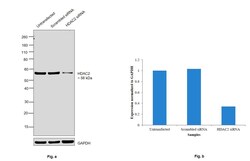
- Knockdown of HDAC2 was achieved by transfecting HEK-293 cells with HDAC2 specific siRNAs (Silencer® select Product # s6494). Western blot analysis (Fig. a) was performed using whole cell extracts from the HDAC2 knockdown cells (Lane 3), non-specific scrambled siRNA transfected cells (Lane 2) and untransfected cells (Lane 1). The blots were probed with Anti-HDAC2 Monoclonal Antibody (3F3) (Product # MA5-18061, 1:1000 dilution) and Goat anti-Mouse IgG (H+L), Superclonal™ Recombinant Secondary Antibody, HRP (Product # A28177, 1:4000 dilution) using the iBright FL 1000 (Product # A32752). Densitometric analysis of this Western Blot is shown in histogram (Fig. b). Decrease in signal upon siRNA mediated knock down confirms that antibody is specific to HDAC2.

- Submitted by
- Invitrogen Antibodies (provider)
- Main image

- Experimental details
- Western blot was performed using Anti-HDAC2 Monoclonal Antibody (3F3) (Product # MA5-18061) and a 56 kDa band corresponding to HDAC2 was observed across the cell lines except MCF 10A. (Fig. a) Modified whole cell extracts (1% SDS) (30 µg lysate) of DU 145 (Lane 1), A549 (Lane 2), PC-3 (Lane 3), Hep G2 (Lane 4), HeLa (Lane 5), HEK-293 (Lane 6), K-562 (Lane 7), SH-SY5Y (Lane 8) and tissue extract (30 µg lysate) of Mouse Thymus; (Fig. b) Modified whole cell extracts (1% SDS) (30 µg lysate) of MCF7 (Lane 1), MDA-MB-231 (Lane 2), SK-BR-3 (Lane 3), T-47D (Lane 4) and MCF 10A were electrophoresed using Novex® NuPAGE® 4-12 % Bis-Tris gel (Product # NP0321BOX). Resolved proteins were then transferred onto a nitrocellulose membrane (Product # IB23001) by iBlot® 2 Dry Blotting System (Product # IB21001). The blot was probed with the primary antibody (1:1000 dilution) and detected by chemiluminescence with Goat anti-Mouse IgG (H+L), Superclonal™ Recombinant Secondary Antibody, HRP (Product # A28177, 1:4000 dilution) using the iBright FL 1000 (Product # A32752). Chemiluminescent detection was performed using Novex® ECL Chemiluminescent Substrate Reagent Kit (Product # WP20005).

Supportive validation
- Submitted by
- Invitrogen Antibodies (provider)
- Main image

- Experimental details
- Immunofluorescence analysis of HDAC2 was performed using 70% confluent log phase HeLa cells. The cells were fixed with 4% paraformaldehyde for 10 minutes, permeabilized with 0.1% Triton™ X-100 for 15 minutes, and blocked with 2% BSA for 1 hour at room temperature. The cells were labeled with HDAC2 Monoclonal Antibody (3F3) (Product MA5-18061) at 2 µg/mL in 0.1% BSA, incubated at 4 degree Celsius overnight and then with Goat anti-Mouse IgG (H+L), Superclonal™ Recombinant Secondary Antibody, Alexa Fluor 488 (Product # A28175) at a dilution of 1:2000 for 45 minutes at room temperature (Panel a: Green). Nuclei (Panel b: Blue) were stained with SlowFade® Gold Antifade Mountant with DAPI (Product # S36938). F-actin (Panel c: Red) was stained with Rhodamine Phalloidin (Product # R415, 1:300). Panel d represents the merged image showing nuclear localization. Panel e represents control cells with no primary antibody to assess background. The images were captured at 60X magnification.

Supportive validation
- Submitted by
- Invitrogen Antibodies (provider)
- Main image

- Experimental details
- Chromatin Immunoprecipitation (ChIP) assay of endogenous HDAC2 protein using Anti-HDAC2 Antibody: ChIP was performed using Anti-HDAC2 Monoclonal Antibody (3F3) (Product # MA5-18061, 2.5 µg) on sheared chromatin from HeLa cells using the MAGnify ChIP System kit (Product # 49-2024). Normal Mouse IgG was used as a negative IP control. The purified DNA was analyzed by qPCR using primers binding to ACTIN and GAPDH transcriptional start site and SAT2 satellite repeats. Data is presented as fold enrichment of the antibody signal versus the negative control IgG using the comparative CT method.
