 Explore
 Validate
 Learn
LearnPA1-886
antibody from Invitrogen Antibodies
Targeting: DMAP1
DNMAP1, DNMTAP1, EAF2, FLJ11543, KIAA1425, MEAF2, SWC4
 Western blot
 Immunohistochemistry
Antibody data
- Antibody Data
- Antigen structure
- References [5]
- Comments [0]
- Validations
- Immunohistochemistry [3]
Submit
Validation data
Reference
Comment
Report error
- Product number
- PA1-886 - Provider product page

- Provider
- Invitrogen Antibodies
- Product name
- DMAP1 Polyclonal Antibody
- Antibody type
- Polyclonal
- Antigen
- Synthetic peptide
- Description
- PA1-886 detects DNA methyltransferase 1 associated protein (DMAP1) from human and mouse cells. PA1-886 has been successfully used in Western blot and Immunohistochemistry (paraffin) procedures. By Western blot, this antibody detects an ~52 kDa band which corresponds to DMAP1 from HeLa cell nuclear extracts. PA1-886 immunizing peptide corresponds to amino acid residues 435-450 from mouse DMAP1. This sequence is completely conserved in human DMAP1. PA1-886 immunizing peptide (Cat. # PEP-118) is available for use in neutralization and control experiments.
- Reactivity
- Human, Mouse
- Host
- Rabbit
- Isotype
- IgG
- Vial size
- 100 μL
- Concentration
- 1 mg/mL
- Storage
- -20°C, Avoid Freeze/Thaw Cycles
Submitted references The mammalian YL1 protein is a shared subunit of the TRRAP/TIP60 histone acetyltransferase and SRCAP complexes.
Structural and functional conservation of the NuA4 histone acetyltransferase complex from yeast to humans.
Structural and functional conservation of the NuA4 histone acetyltransferase complex from yeast to humans.
RGS6 interacts with DMAP1 and DNMT1 and inhibits DMAP1 transcriptional repressor activity.
Identification of new subunits of the multiprotein mammalian TRRAP/TIP60-containing histone acetyltransferase complex.
Cai Y, Jin J, Florens L, Swanson SK, Kusch T, Li B, Workman JL, Washburn MP, Conaway RC, Conaway JW
The Journal of biological chemistry 2005 Apr 8;280(14):13665-70
The Journal of biological chemistry 2005 Apr 8;280(14):13665-70
Structural and functional conservation of the NuA4 histone acetyltransferase complex from yeast to humans.
Doyon Y, Selleck W, Lane WS, Tan S, Côté J
Molecular and cellular biology 2004 Mar;24(5):1884-96
Molecular and cellular biology 2004 Mar;24(5):1884-96
Structural and functional conservation of the NuA4 histone acetyltransferase complex from yeast to humans.
Doyon Y, Selleck W, Lane WS, Tan S, Côté J
Molecular and cellular biology 2004 Mar;24(5):1884-96
Molecular and cellular biology 2004 Mar;24(5):1884-96
RGS6 interacts with DMAP1 and DNMT1 and inhibits DMAP1 transcriptional repressor activity.
Liu Z, Fisher RA
The Journal of biological chemistry 2004 Apr 2;279(14):14120-8
The Journal of biological chemistry 2004 Apr 2;279(14):14120-8
Identification of new subunits of the multiprotein mammalian TRRAP/TIP60-containing histone acetyltransferase complex.
Cai Y, Jin J, Tomomori-Sato C, Sato S, Sorokina I, Parmely TJ, Conaway RC, Conaway JW
The Journal of biological chemistry 2003 Oct 31;278(44):42733-6
The Journal of biological chemistry 2003 Oct 31;278(44):42733-6
No comments: Submit comment
Supportive validation
- Submitted by
- Invitrogen Antibodies (provider)
- Main image

- Experimental details
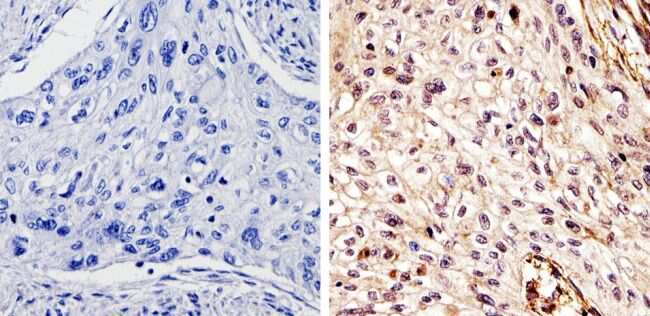
- Immunohistochemistry analysis of DMAP1 showing staining in the cytoplasm and nucleus of paraffin-treated human esophageal carcinoma (right) compared with a negative control in the absence of primary antibody (left). To expose target proteins, antigen retrieval was performed using 10mM sodium citrate (pH 6.0), microwaved for 8-15 min. Following antigen retrieval, tissues were blocked in 3% H2O2-methanol for 15 min at room temperature, washed with ddH2O and PBS, and then probed with a DMAP1 polyclonal antibody (Product # PA1-886) diluted by 3% BSA-PBS at a dilution of 1:100 overnight at 4°C in a humidified chamber. Tissues were washed extensively in PBST and detection was performed using an HRP-conjugated secondary antibody followed by colorimetric detection using a DAB kit. Tissues were counterstained with hematoxylin and dehydrated with ethanol and xylene to prep for mounting.
- Submitted by
- Invitrogen Antibodies (provider)
- Main image
- Experimental details
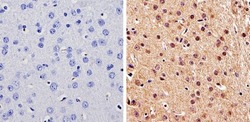
- Immunohistochemistry analysis of DMAP1 showing staining in the cytoplasm and nucleus of paraffin-treated mouse brain tissue (right) compared with a negative control in the absence of primary antibody (left). To expose target proteins, antigen retrieval was performed using 10mM sodium citrate (pH 6.0), microwaved for 8-15 min. Following antigen retrieval, tissues were blocked in 3% H2O2-methanol for 15 min at room temperature, washed with ddH2O and PBS, and then probed with a DMAP1 polyclonal antibody (Product # PA1-886) diluted by 3% BSA-PBS at a dilution of 1:100 overnight at 4°C in a humidified chamber. Tissues were washed extensively in PBST and detection was performed using an HRP-conjugated secondary antibody followed by colorimetric detection using a DAB kit. Tissues were counterstained with hematoxylin and dehydrated with ethanol and xylene to prep for mounting.
- Submitted by
- Invitrogen Antibodies (provider)
- Main image
- Experimental details
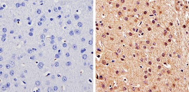
- Immunohistochemistry analysis of DMAP1 showing staining in the cytoplasm and nucleus of paraffin-treated mouse uterus tissue (right) compared with a negative control in the absence of primary antibody (left). To expose target proteins, antigen retrieval was performed using 10mM sodium citrate (pH 6.0), microwaved for 8-15 min. Following antigen retrieval, tissues were blocked in 3% H2O2-methanol for 15 min at room temperature, washed with ddH2O and PBS, and then probed with a DMAP1 polyclonal antibody (Product # PA1-886) diluted by 3% BSA-PBS at a dilution of 1:100 overnight at 4°C in a humidified chamber. Tissues were washed extensively in PBST and detection was performed using an HRP-conjugated secondary antibody followed by colorimetric detection using a DAB kit. Tissues were counterstained with hematoxylin and dehydrated with ethanol and xylene to prep for mounting.
