Explore
 Validate
 Learn
Learn Western blot
 Immunocytochemistry
Antibody data
- Antibody Data
- Antigen structure
- References [1]
- Comments [0]
- Validations
- Immunocytochemistry [1]
- Other assay [2]
Submit
Validation data
Reference
Comment
Report error
- Product number
- PA5-77078 - Provider product page

- Provider
- Invitrogen Antibodies
- Product name
- RAD51C Polyclonal Antibody
- Antibody type
- Polyclonal
- Antigen
- Recombinant full-length protein
- Description
- The antibody was affinity-purified from rabbit antiserum by affinity-chromatography using epitope-specific immunogen and the purity is > 95% (by SDS-PAGE).
- Reactivity
- Human, Mouse
- Host
- Rabbit
- Isotype
- IgG
- Vial size
- 100 μL
- Concentration
- 1 mg/mL
- Storage
- Store at 4°C short term. For long term storage, store at -20°C, avoiding freeze/thaw cycles.
Submitted references LncRNA CTBP1-DT-encoded microprotein DDUP sustains DNA damage response signalling to trigger dual DNA repair mechanisms.
Yu R, Hu Y, Zhang S, Li X, Tang M, Yang M, Wu X, Li Z, Liao X, Xu Y, Li M, Chen S, Qian W, Gong LY, Song L, Li J
Nucleic acids research 2022 Aug 12;50(14):8060-8079
Nucleic acids research 2022 Aug 12;50(14):8060-8079
No comments: Submit comment
Supportive validation
- Submitted by
- Invitrogen Antibodies (provider)
- Main image
- Experimental details
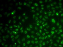
- Immunofluorescence analysis of RAD51C in U20S cells. Samples were incubated with RAD51C polyclonal antibody (Product # PA5-77078).

Supportive validation
- Submitted by
- Invitrogen Antibodies (provider)
- Main image
- Experimental details
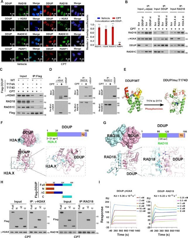
- Figure 5. Phosphorylated DDUP forms a complex with gamma-H2AX and RAD18. ( A ) IF staining analysis of the co-localisation of DDUP foci with RAD18 foci, gamma-H2AX foci, RAD51C-foci and PARP1-foci in vehicle- and CPT (10 muM, 1 h)-treated HeLa cells. Scale bar = 5 mum. Co-localization the fluorescence between molecules was quantified using the Manders' overlap coefficients algorithm. ( B ) Co-IP assay analysis of the formation of the DDUP/gamma-H2AX/RAD18/RAD51C complex in the indicated gene-silenced 293T cells treated with CPT (10 muM) for 1 h. ( C ) Co-IP assay analysis of the interaction of WT and mutated DDUP with gamma-H2AX, RAD18 and RAD51C in CPT (10 muM, 1 h) or without CPT-treated 293T cells. ( D ) Far-western blotting analysis of the direct interaction of DDUP/gamma-H2AX using anti-gamma-H2AX antibody-immunoprecipitated proteins (left), or DDUP/RAD18 using anti-RAD18 antibody-immunoprecipitated proteins (middle), or DDUP/RAD51C using anti-RAD51C antibody-immunoprecipitated proteins in RAD18-silenced cells (right), then detected using anti-DDUP antibody. Recombinant DDUP/T174D protein served as control. ( E ) Left: the 3D structure of WT DDUP in the dense state obtained from the I-TASSER server. Right: the 3D structure of the DDUP mutant (T174 to D174), which mimics phosphorylation of DDUP, in the loose state obtained from the I-TASSER server. ( F ) Molecular docking of H2A.X and DDUP. The 3D structure of H2A.X obtained from the I-TASSER server. The combined surface,
- Submitted by
- Invitrogen Antibodies (provider)
- Main image
- Experimental details
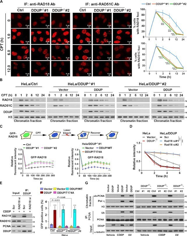
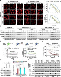
- Figure 6. DDUP enhances the retention of RAD18 at DNA damage sites. ( A ) Representative images (left) and time course (right) of the formation of CPT (10 muM)-induced RAD18 and RAD51C foci in control and DDUP-KO HeLa cells and allowed to recover for the indicated times. The RAD18- and RAD51C foci was examined every 10 min in the CPT-treated cells within the first 2 h. Cells containing more than 10 RAD18 and RAD51C foci per nucleus were scored. ( B ) Chromatin fraction and IB analysis of DNA-bound RAD18, RAD51C and DDUP in the indicated CPT (10 muM)-treated cells and allowed to recover for the indicated times. H3 served as a loading control. ( C ) Quantitative FRAP analysis of GFP-RAD18 in GFP-RAD18-transfected control and DDUP-KO HeLa cells (right), and in DDUP-KO HeLa cells co-transfected with GFP-RAD18 and vector, GFP-RAD18, and DDUP/WT, or GFP-RAD18 and DDUP/T174A, treated with CPT (10 muM) and allowed to recover for the indicated times. ( D ) Kinetics of gamma-H2AX signals in the indicated cells in response to laser micro-irradiation and allowed to recover for the indicated times ( n = 100). ( E ) IP assay analysis of the DDUP/RAD51C and DDUP/PCNA interaction in control and RAD18-silenced 293T cells treated with CDDP (5 muM, 1 h). ( F ) Homologous recombination repair assays performed in the indicated cells. ( G ) IP/IB analysis of the regulatory effect of DDUP dysregulation on PCNA monoubiquitination in the indicated cells treated with CDDP (5 muM, 1 h) or UV radiation