 Explore
 Validate
 Learn
Learn ELISA
ELISA Immunocytochemistry
 Immunoprecipitation
Antibody data
- Antibody Data
- Antigen structure
- References [0]
- Comments [0]
- Validations
- Immunocytochemistry [2]
- Other assay [1]
Submit
Validation data
Reference
Comment
Report error
- Product number
- MA5-14736 - Provider product page

- Provider
- Invitrogen Antibodies
- Product name
- Ferritin Monoclonal Antibody (513C10)
- Antibody type
- Monoclonal
- Antigen
- Other
- Description
- MA5-14736 detects Ferritin in ELISA, IP, IF, and RIA applications and shows reactivity to human samples. The MA5-14736 Immunogen is human liver ferritin. MA5-14736 detects ferritin which has a predicted molecular weight of approximately 19kDa. Product MA514736 is a smaller package size of MIF2504 (formerly sold as a Seradyn product).
- Reactivity
- Human
- Host
- Mouse
- Isotype
- IgG
- Antibody clone number
- 513C10
- Vial size
- 100 µg
- Concentration
- 1 mg/mL
- Storage
- Maintain refrigerated at 2-8°C for up to 6 months. For long term storage store at -20°C
No comments: Submit comment
Supportive validation
- Submitted by
- Invitrogen Antibodies (provider)
- Main image
- Experimental details
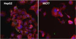
- Immunofluorescent analysis of Ferritin (FTL, green) in HepG2 cells and MCF7 cells. The cells were fixed with formalin for 15 minutes, permeabilized with 0.1% Triton X-100 in TBS for 10 minutes, and blocked with 1% Blocker BSA (Product # 37525) for 15 minutes at room temperature. Cells were stained with a Ferritin monoclonal antibody (Product # MA5-14736), at a dilution of 1:200 for at least 1 hour at room temperature, and then incubated with a DyLight 488 goat anti-mouse IgG secondary antibody (Product # 35503) at a dilution of 1:500 for 30 minutes at room temperature (both panels, green). F-Actin (both panels, red) was stained with DyLight 554 Phalloidin (Product # 21834) and nuclei (both panels, blue) were stained with Hoechst 33342 dye (Product # 62249). Images were taken on a Thermo Scientific ToxInsight at 20X magnification.
- Submitted by
- Invitrogen Antibodies (provider)
- Main image
- Experimental details
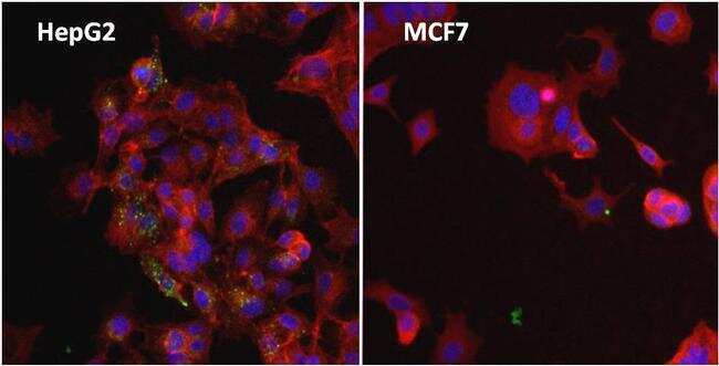
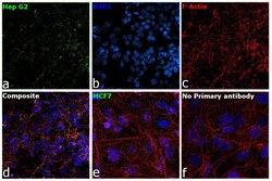
- Immunofluorescence analysis of Ferritin was performed using 70% confluent log phase Hep G2 and MCF7 cells. The cells were fixed with 4% paraformaldehyde for 10 minutes, permeabilized with 0.1% Triton™ X-100 for 15 minutes, and blocked with 2% BSA for 45 minutes at room temperature. The cells were labeled with Ferritin Monoclonal Antibody (513C10) (Product # MA5-14736) at 1:100 dilution in 0.1% BSA, incubated at 4 degree celsius overnight and then labeled with Donkey anti-Mouse IgG (H+L) Highly Cross-Adsorbed Secondary Antibody, Alexa Fluor Plus 488 (Product # A32766), (1:2000 dilution), for 45 minutes at room temperature (Panel a: Green). Nuclei (Panel b:Blue) were stained with ProLong™ Diamond Antifade Mountant with DAPI (Product # P36962). F-actin (Panel c: Red) was stained with Rhodamine Phalloidin (Product # R415, 1:300). Panel d represents the merged image showing cytoplasmic localization. Panel e represents MCF7 cells with reduced expression. Panel f represents control cells with no primary antibody to assess background. The images were captured at 60X magnification.

Supportive validation
- Submitted by
- Invitrogen Antibodies (provider)
- Main image

- Experimental details
- Direct ELISA analysis of Ferritin was performed by coating wells of a 96-well plate with 100 µL per well of Human Ferritin or BSA diluted in carbonate/bicarbonate buffer (Product # 28382), starting at a concentration of 1 µg/mL and serially diluting 2-fold to a concentration of 2 ng/mL, overnight at 4C. Wells of the plate were washed, blocked with StartingBlock blocking buffer (Product # 37538), and incubated with 100 µL per well of a mouse anti-ferritin monoclonal antibody (Product # MA5-14736) at a concentration of 1 µg/mL for 1 hour at room temperature. The plate was washed, then incubated with 100 µL per well of an HRP-conjugated goat anti-mouse IgG secondary antibody (Product # 31430) at a dilution of 1:25000 for 30 minutes at room temperature. Detection was performed using 1-Step Ultra TMB substrate (Product # 34028) for 10 minutes at room temperature in the dark. The reaction was stopped with 0.16M sulfuric acid, and absorbances were read on a spectrophotometer at 450-550 nm.
