 Explore
 Validate
 Learn
Learn Western blot
Antibody data
- Antibody Data
- Antigen structure
- References [1]
- Comments [0]
- Validations
- Western blot [1]
- ELISA [1]
- Immunohistochemistry [1]
Submit
Validation data
Reference
Comment
Report error
- Product number
- PA5-19059 - Provider product page

- Provider
- Invitrogen Antibodies
- Product name
- Ferritin Light Chain Polyclonal Antibody
- Antibody type
- Polyclonal
- Antigen
- Synthetic peptide
- Description
- This antibody is predicted to react with bovine, canine, mouse and rat based on sequence homology. This antibody is tested in Peptide ELISA: antibody detection limit dilution 32,000.
- Reactivity
- Human, Mouse, Rat
- Host
- Goat
- Isotype
- IgG
- Vial size
- 100 µg
- Concentration
- 0.5 mg/mL
- Storage
- -20° C, Avoid Freeze/Thaw Cycles
Submitted references Neuropathology of sporadic Parkinson disease before the appearance of parkinsonism: preclinical Parkinson disease.
Ferrer I, Martinez A, Blanco R, Dalfó E, Carmona M
Journal of neural transmission (Vienna, Austria : 1996) 2011 May;118(5):821-39
Journal of neural transmission (Vienna, Austria : 1996) 2011 May;118(5):821-39
No comments: Submit comment
Supportive validation
- Submitted by
- Invitrogen Antibodies (provider)
- Main image
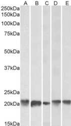
- Experimental details
- Western blot analysis of Ferritin using a Ferritin polyclonal antibody (Product # PA5-19059) at a concentration of 0.3 µg/mL. Human Brain (Cerebellum) (A), Liver (B), Human Placenta (C) 22kDa in MBR (D), RBR (E) lysate (35 µg protein in RIPA buffer). Primary incubation was 1 hour. Detected by chemiluminescence.

Supportive validation
- Submitted by
- Invitrogen Antibodies (provider)
- Main image

- Experimental details
- Elisa analysis of ferritin using a ferritin polyclonal antibody (Product # PA5-19059) at a concentration of 1.5 µg/mL.

Supportive validation
- Submitted by
- Invitrogen Antibodies (provider)
- Main image
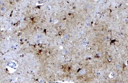
- Experimental details
- Immunohistochemichal analysis of Ferritin in Human Brain Cortex using a Ferritin polyclonal antibody (Product # PA5-19059) at a concentration of 3.8 µg/mL. The sample was paraffin embedded, and heat treated antigen retrieval was used to detect the target with HRP staining.
