 Explore
 Validate
 Learn
Learn Western blot
 ELISA
ELISAAntibody data
- Antibody Data
- Antigen structure
- References [1]
- Comments [0]
- Validations
- Western blot [3]
- Immunohistochemistry [1]
- Other assay [1]
Submit
Validation data
Reference
Comment
Report error
- Product number
- PA5-29599 - Provider product page

- Provider
- Invitrogen Antibodies
- Product name
- Ferritin Light Chain Polyclonal Antibody
- Antibody type
- Polyclonal
- Antigen
- Recombinant protein fragment
- Description
- Recommended positive controls: HepG2, mouse liver. Predicted reactivity: Dog (87%), Cat (86%), Pig (88%), Rabbit (86%), Rhesus Monkey (97%), Bovine (84%), Guinea pig (84%). Store product as a concentrated solution. Centrifuge briefly prior to opening the vial.
- Reactivity
- Human, Mouse, Rat
- Host
- Rabbit
- Isotype
- IgG
- Vial size
- 100 µL
- Concentration
- 1 mg/mL
- Storage
- Store at 4°C short term. For long term storage, store at -20°C, avoiding freeze/thaw cycles.
Submitted references Enhanced Cellular Uptake of H-Chain Human Ferritin Containing Gold Nanoparticles.
Moglia I, Santiago M, Guerrero S, Soler M, Olivera-Nappa A, Kogan MJ
Pharmaceutics 2021 Nov 19;13(11)
Pharmaceutics 2021 Nov 19;13(11)
No comments: Submit comment
Supportive validation
- Submitted by
- Invitrogen Antibodies (provider)
- Main image

- Experimental details
- Western Blot analysis of Ferritin Light Chain was performed by separating 50 µg of mouse tissue extract by 12% SDS-PAGE. Proteins were transferred to a membrane and probed with a Ferritin Light Chain Polyclonal Antibody (Product # PA5-29599) at a dilution of 1:1000.

- Submitted by
- Invitrogen Antibodies (provider)
- Main image

- Experimental details
- Western Blot using Ferritin Light Chain Polyclonal Antibody (Product # PA5-29599). Sample (30 µg of whole cell lysate). Lane A: Hep G2. 12% SDS PAGE. Ferritin Light Chain Polyclonal Antibody (Product # PA5-29599) diluted at 1:1,000.

- Submitted by
- Invitrogen Antibodies (provider)
- Main image
- Experimental details
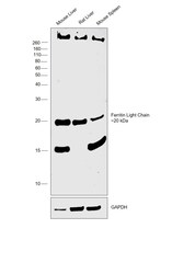
- Western blot was performed using Anti-Ferritin Light Chain Polyclonal Antibody (Product # PA5-29599) and a 20kDa band corresponding to Ferritin Light Chain was observed across tissues tested with lower levels in mouse spleen. Tissue extracts (30 µg lysate) of Mouse Liver (Lane 1), Rat Liver (Lane 2), Mouse Spleen (Lane 3) were electrophoresed using NuPAGE™ 12% Bis-Tris Protein Gel (Product # NP0341BOX). Resolved proteins were then transferred onto a Nitrocellulose membrane (Product # IB23001) by iBlot® 2 Dry Blotting System (Product # IB21001). The blot was probed with the primary antibody (1:1000 dilution) and detected by chemiluminescence with Goat anti-Rabbit IgG (H+L) Superclonal™ Recombinant Secondary Antibody, HRP (Product # A27036,1:4000 dilution) using the iBright FL 1000 (Product # A32752). Chemiluminescent detection was performed using Novex® ECL Chemiluminescent Substrate Reagent Kit (Product # WP20005). Ferritin is a holoenzyme shell with a molecular weight of about 450 kDa, and consists of 24 subunits of two types, H(heavy) and L(light), hence band above 260kDa could correspond to the holoenzyme shell of 450kDa.

Supportive validation
- Submitted by
- Invitrogen Antibodies (provider)
- Main image
- Experimental details
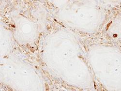
- Immunohistochemical analysis of paraffin-embedded Ca922 xenograft, using Ferritin Light Chain (Product # PA5-29599) antibody at 1:100 dilution. Antigen Retrieval: EDTA based buffer, pH 8.0, 15 min.

Supportive validation
- Submitted by
- Invitrogen Antibodies (provider)
- Main image

- Experimental details
- Figure 6 Confocal microscopy results for different cell lines after incubation with FT-AuNP. ( a ) Confocal images of HEK293T, CH3/10T1/2, BEND3, and HT29 were obtained after incubation for 24 h with H-ferritin (FTH) and L-ferritin (FTL), with or without AuNP or iron oxides (anti FTH or FTL in red; (DAPI) nucleus in blue; scale bars: 20 mum). ( b ) Plots representing fold changes in MFI (mean fluorescence intensity) +- SD from more than three independent experiments.
