Explore
 Validate
 Learn
Learn Western blot
Antibody data
- Antibody Data
- Antigen structure
- References [0]
- Comments [0]
- Validations
- Western blot [1]
- Immunohistochemistry [2]
- Flow cytometry [1]
Submit
Validation data
Reference
Comment
Report error
- Product number
- TA303540 - Provider product page

- Provider
- OriGene
- Product name
- Rabbit monoclonal antibody against HSF1 Phospho (pS326) (EP1713Y ) (phospho-specific)
- Antibody type
- Monoclonal
- Description
- Rabbit monoclonal antibody against HSF1 Phospho (pS326) (EP1713Y ) (phospho-specific)
- Host
- Rabbit
- Conjugate
- Unconjugated
- Epitope
- HSF1
- Isotype
- IgG
- Antibody clone number
- EP1713Y
- Vial size
- 100 µl
- Concentration
- NULL
No comments: Submit comment
Supportive validation
- Submitted by
- OriGene (provider)
- Main image

- Experimental details
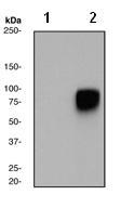
- Western blot - HSF1 (phospho S326) antibody [EP1713Y]; All lanes : Anti-HSF1 (phospho S326) antibody [EP1713Y] at 1/10000 dilution.Lane 1 : HeLa cell lysates (untreated).Lane 2 : HeLa cell lysates, treated with heat (44o C).Lysates/proteins at 10 ug per lane.Secondary.Goat anti-rabbit HRP at 1/1000 dilution.Predicted band size : 57 kDa.Observed band size : 82 kDa .
- Validation comment
- WB
Supportive validation
- Submitted by
- OriGene (provider)
- Main image

- Experimental details
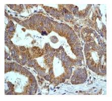
- Immunohistochemistry (Formalin/PFA-fixed paraffin-embedded sections) - HSF1 (phospho S326) antibody [EP1713Y]; Immunohistochemical analysis of paraffin-embedded human stomach using TA303540, at 1/100 dilution.
- Validation comment
- IHC
- Submitted by
- OriGene (provider)
- Main image

- Experimental details
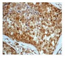
- Immunohistochemistry (Formalin/PFA-fixed paraffin-embedded sections) - HSF1 (phospho S326) antibody [EP1713Y]; Immunohistochemical analysis of paraffin-embedded human breast carcinoma, using TA303540, at 1/100 dilution.
- Validation comment
- IHC
Supportive validation
- Submitted by
- OriGene (provider)
- Main image
- Experimental details
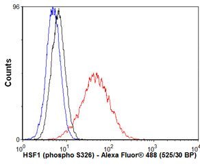
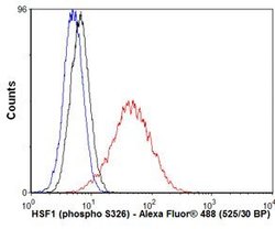
- Flow Cytometry - Anti-HSF1 (phospho S326) antibody; Overlay histogram showing HeLa cells stained with TA303540 (red line). The secondary antibody used was Alexa Fluor 488 goat anti-rabbit IgG (H+L) at 1:2000. Isotype control antibody (black line) was rabbit IgG (monoclonal) used under the same conditions. Unlabelled sample (blue line) was also used as a control. This antibody gave a positive signal in HeLa cells under the same conditions.
- Validation comment
- FC