Explore
 Validate
 Learn
Learn Western blot
 Immunocytochemistry
Antibody data
- Antibody Data
- Antigen structure
- References [1]
- Comments [0]
- Validations
- Immunocytochemistry [1]
- Immunohistochemistry [1]
Submit
Validation data
Reference
Comment
Report error
- Product number
- GTX22861 - Provider product page

- Provider
- GeneTex
- Proper citation
- GeneTex Cat#GTX22861, RRID:AB_384881
- Product name
- SERCA2 ATPase antibody [2A7-A1]
- Antibody type
- Monoclonal
- Reactivity
- Human, Mouse, Rat, Canine, Drosophila, Guinea Pig, Porcine, Rabbit, Sheep, Xenopus
- Host
- Mouse
Submitted references Targeted disruption of PDE3B, but not PDE3A, protects murine heart from ischemia/reperfusion injury.
Chung YW, Lagranha C, Chen Y, Sun J, Tong G, Hockman SC, Ahmad F, Esfahani SG, Bae DH, Polidovitch N, Wu J, Rhee DK, Lee BS, Gucek M, Daniels MP, Brantner CA, Backx PH, Murphy E, Manganiello VC
Proceedings of the National Academy of Sciences of the United States of America 2015 Apr 28;112(17):E2253-62
Proceedings of the National Academy of Sciences of the United States of America 2015 Apr 28;112(17):E2253-62
No comments: Submit comment
Supportive validation
- Submitted by
- GeneTex (provider)
- Main image

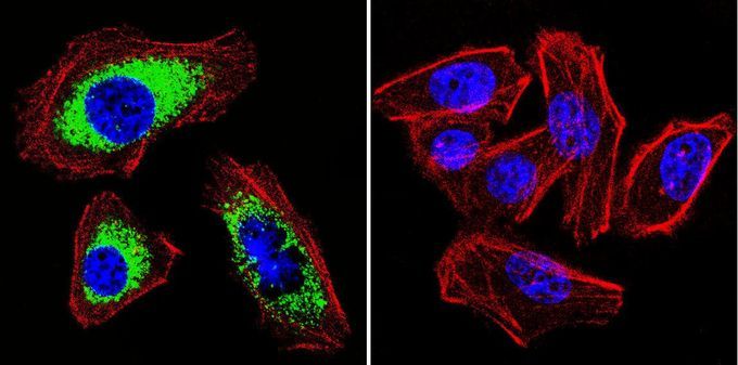
- Experimental details
- Immunofluorescent analysis of SERCA2 ATPase in A549 cells. SERCA2 ATPase staining (green), F-Actin staining with Phalloidin (red) and nuclei with DAPI (blue) is shown. Cells were grown on slides and fixed with formaldehyde prior to staining. Cells were probed without (control) or with SERCA2 ATPase antibody [2A7-A1] at a dilution of 1:100-1:200 over night at 4¢XC, washed with PBS and incubated with a proper secondary antibody. Images were taken at 60X magnification.
Supportive validation
- Submitted by
- GeneTex (provider)
- Main image
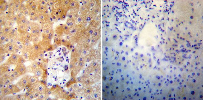
- Experimental details
- Immunohistochemistry was performed on normal deparaffinized human liver tissue tissues. To expose target proteins, heat induced antigen retrieval was performed using 10mM sodium citrate (pH6.0) buffer, microwaved for 8-15 minutes. Following antigen retrieval tissues were blocked in 3% BSA-PBS for 30 minutes at room temperature. Tissues were then probed at a dilution of 1:100 with without SERCA2 ATPase antibody [2A7-A1] overnight at 4¢XC in a humidified chamber. Tissues were washed extensively with PBST and endogenous peroxidase activity was quenched with a peroxidase suppressor. Detection was performed using a biotin-conjugated secondary antibody and SA-HRP, followed by colorimetric detection using DAB. Tissues were counterstained with hematoxylin and prepped for mounting.