 Explore
 Validate
 Learn
Learn Western blot
 ELISA
ELISAAntibody data
- Antibody Data
- Antigen structure
- References [10]
- Comments [0]
- Validations
- Western blot [3]
- Immunohistochemistry [1]
Submit
Validation data
Reference
Comment
Report error
- Product number
- NB110-55376 - Provider product page

- Provider
- Novus Biologicals
- Proper citation
- Novus Cat#NB110-55376, RRID:AB_838957
- Product name
- Rabbit Polyclonal TDP-43/TARDBP Antibody
- Antibody type
- Polyclonal
- Description
- Immunogen affinity purified.
- Reactivity
- Human, Mouse, Chicken/Avian, Simian, Xenopus, Zebrafish
- Host
- Rabbit
- Isotype
- IgG
- Vial size
- 0.1 ml
- Concentration
- 1 mg/ml
- Storage
- Store at 4C. Do not freeze.
Submitted references The cleavage pattern of TDP-43 determines its rate of clearance and cytotoxicity.
TDP-43 as a possible biomarker for frontotemporal lobar degeneration: a systematic review of existing antibodies.
Full-length TDP-43 forms toxic amyloid oligomers that are present in frontotemporal lobar dementia-TDP patients.
Prion-like nuclear aggregation of TDP-43 during heat shock is regulated by HSP40/70 chaperones.
Loss of ALS-associated TDP-43 in zebrafish causes muscle degeneration, vascular dysfunction, and reduced motor neuron axon outgrowth.
TDP-43 promotes microRNA biogenesis as a component of the Drosha and Dicer complexes.
Downregulation of VAPB expression in motor neurons derived from induced pluripotent stem cells of ALS8 patients.
TDP-43 dimerizes in human cells in culture.
TDP-43 mutant transgenic mice develop features of ALS and frontotemporal lobar degeneration.
Evidence of multisystem disorder in whole-brain map of pathological TDP-43 in amyotrophic lateral sclerosis.
Li Q, Yokoshi M, Okada H, Kawahara Y
Nature communications 2015 Jan 29;6:6183
Nature communications 2015 Jan 29;6:6183
TDP-43 as a possible biomarker for frontotemporal lobar degeneration: a systematic review of existing antibodies.
Goossens J, Vanmechelen E, Trojanowski JQ, Lee VM, Van Broeckhoven C, van der Zee J, Engelborghs S
Acta neuropathologica communications 2015 Apr 1;3:15
Acta neuropathologica communications 2015 Apr 1;3:15
Full-length TDP-43 forms toxic amyloid oligomers that are present in frontotemporal lobar dementia-TDP patients.
Fang YS, Tsai KJ, Chang YJ, Kao P, Woods R, Kuo PH, Wu CC, Liao JY, Chou SC, Lin V, Jin LW, Yuan HS, Cheng IH, Tu PH, Chen YR
Nature communications 2014 Sep 12;5:4824
Nature communications 2014 Sep 12;5:4824
Prion-like nuclear aggregation of TDP-43 during heat shock is regulated by HSP40/70 chaperones.
Udan-Johns M, Bengoechea R, Bell S, Shao J, Diamond MI, True HL, Weihl CC, Baloh RH
Human molecular genetics 2014 Jan 1;23(1):157-70
Human molecular genetics 2014 Jan 1;23(1):157-70
Loss of ALS-associated TDP-43 in zebrafish causes muscle degeneration, vascular dysfunction, and reduced motor neuron axon outgrowth.
Schmid B, Hruscha A, Hogl S, Banzhaf-Strathmann J, Strecker K, van der Zee J, Teucke M, Eimer S, Hegermann J, Kittelmann M, Kremmer E, Cruts M, Solchenberger B, Hasenkamp L, van Bebber F, Van Broeckhoven C, Edbauer D, Lichtenthaler SF, Haass C
Proceedings of the National Academy of Sciences of the United States of America 2013 Mar 26;110(13):4986-91
Proceedings of the National Academy of Sciences of the United States of America 2013 Mar 26;110(13):4986-91
TDP-43 promotes microRNA biogenesis as a component of the Drosha and Dicer complexes.
Kawahara Y, Mieda-Sato A
Proceedings of the National Academy of Sciences of the United States of America 2012 Feb 28;109(9):3347-52
Proceedings of the National Academy of Sciences of the United States of America 2012 Feb 28;109(9):3347-52
Downregulation of VAPB expression in motor neurons derived from induced pluripotent stem cells of ALS8 patients.
Mitne-Neto M, Machado-Costa M, Marchetto MC, Bengtson MH, Joazeiro CA, Tsuda H, Bellen HJ, Silva HC, Oliveira AS, Lazar M, Muotri AR, Zatz M
Human molecular genetics 2011 Sep 15;20(18):3642-52
Human molecular genetics 2011 Sep 15;20(18):3642-52
TDP-43 dimerizes in human cells in culture.
Shiina Y, Arima K, Tabunoki H, Satoh J
Cellular and molecular neurobiology 2010 May;30(4):641-52
Cellular and molecular neurobiology 2010 May;30(4):641-52
TDP-43 mutant transgenic mice develop features of ALS and frontotemporal lobar degeneration.
Wegorzewska I, Bell S, Cairns NJ, Miller TM, Baloh RH
Proceedings of the National Academy of Sciences of the United States of America 2009 Nov 3;106(44):18809-14
Proceedings of the National Academy of Sciences of the United States of America 2009 Nov 3;106(44):18809-14
Evidence of multisystem disorder in whole-brain map of pathological TDP-43 in amyotrophic lateral sclerosis.
Geser F, Brandmeir NJ, Kwong LK, Martinez-Lage M, Elman L, McCluskey L, Xie SX, Lee VM, Trojanowski JQ
Archives of neurology 2008 May;65(5):636-41
Archives of neurology 2008 May;65(5):636-41
No comments: Submit comment
Supportive validation
- Submitted by
- Novus Biologicals (provider)
- Main image

- Experimental details
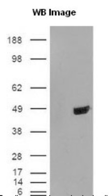
- Western Blot: TDP-43/TARDBP Antibody [NB110-55376] - Cells were transfected with the pCMV6-ENTRY control or pCMV6-ENTRY TARDBP cDNA for 48 hrs and lysed. Equivalent amounts of cell lysates (5 ug per lane) were separated by SDS-PAGE and immunoblotted with anti-TARDBP.
- Submitted by
- Novus Biologicals (provider)
- Main image

- Experimental details
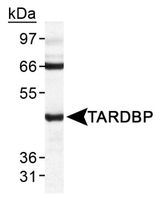
- Western Blot: TDP-43/TARDBP Antibody [NB110-55376] - TARDBP antibody was tested in HeLa WCE.
- Submitted by
- Novus Biologicals (provider)
- Main image
- Experimental details
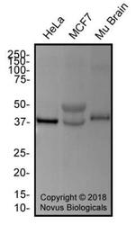
- Western Blot: TDP-43/TARDBP Antibody [NB110-55376] - Total protein from HeLa, MCF7 and mouse brain was separated on a 12% gel by SDS-PAGE, transferred to PVDF membrane and blocked in 5% non-fat milk in TBST. The membrane was probed with 1.0 ug/ml anti-TARDBP in 1% block buffer and detected with an anti-rabbit HRP secondary antibody using chemiluminescence.
Supportive validation
- Submitted by
- Novus Biologicals (provider)
- Main image
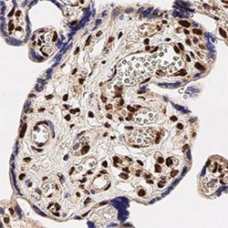
- Experimental details
- Immunohistochemistry-Paraffin: TDP-43/TARDBP Antibody [NB110-55376] - Analysis of FFPE human placenta using TDP-43 antibody at 1:250 on a Bond Rx autostainer (Leica Biosystems). The assay involved 20 minutes of heat induced antigen retrieval (HIER) using 10mM sodium citrate buffer (pH 6.0) and endogenous peroxidase quenching with peroxide block. The sections were incubated with primary antibody for 30 minutes and Bond Polymer Refine Detection (Leica Biosystems) with DAB was used for signal development followed by counterstaining with hematoxylin. Whole slide scanning and capturing of representative images was performed using Aperio AT2 (Leica Biosystems). Nuclear staining was observed. Staining was performed by Histowiz.
