 Explore
 Validate
 Learn
LearnMA5-35273
antibody from Invitrogen Antibodies
Targeting: TARDBP
ALS10, TDP-43
 Western blot
 ELISA
ELISA Immunocytochemistry Immunoprecipitation Immunohistochemistry Chromatin Immunoprecipitation Other assay
Antibody data
- Antibody Data
- Antigen structure
- References [1]
- Comments [0]
- Validations
- Immunocytochemistry [6]
- Immunohistochemistry [3]
- Chromatin Immunoprecipitation [1]
- Other assay [1]
Submit
Validation data
Reference
Comment
Report error
- Product number
- MA5-35273 - Provider product page

- Provider
- Invitrogen Antibodies
- Product name
- TDP-43 Recombinant Rabbit Monoclonal Antibody (4R5L7)
- Antibody type
- Monoclonal
- Antigen
- Synthetic peptide
- Description
- Immunogen sequence: GNNQGSNMGG GMNFGAFSIN PAMMAAAQAA LQSSWGMMGM LASQQNQSGP SGNNQNQGNM QREPNQAFGS GNNSYSGSNS GAAIGWGSAS NAGSGSGFNG G
- Reactivity
- Human, Mouse, Rat
- Host
- Rabbit
- Isotype
- IgG
- Antibody clone number
- 4R5L7
- Vial size
- 100 μL
- Concentration
- 0.8 mg/mL
- Storage
- -20°C, Avoid Freeze/Thaw Cycles
Submitted references Importin-Mediated Pathological Tau Nuclear Translocation Causes Disruption of the Nuclear Lamina, TDP-43 Mislocalization and Cell Death.
Candia RF, Cohen LS, Morozova V, Corbo C, Alonso AD
Frontiers in molecular neuroscience 2022;15:888420
Frontiers in molecular neuroscience 2022;15:888420
No comments: Submit comment
Supportive validation
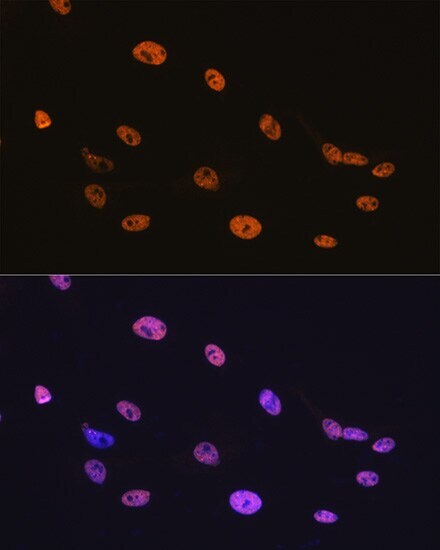
- Submitted by
- Invitrogen Antibodies (provider)
- Main image
- Experimental details
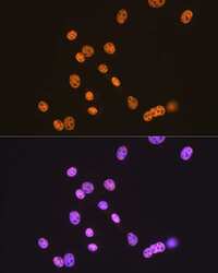
- Immunocytochemical analysis of TDP-43 performed on C6 cells using a TDP-43 monoclonal antibody (Product # MA5-35273) at a dilution of 1:100. An HRP Goat Anti-Rabbit IgG (H+L) secondary antibody was applied at a dilution of 1:10,000.
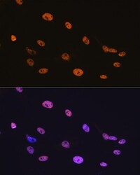
- Submitted by
- Invitrogen Antibodies (provider)
- Main image
- Experimental details
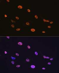
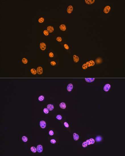
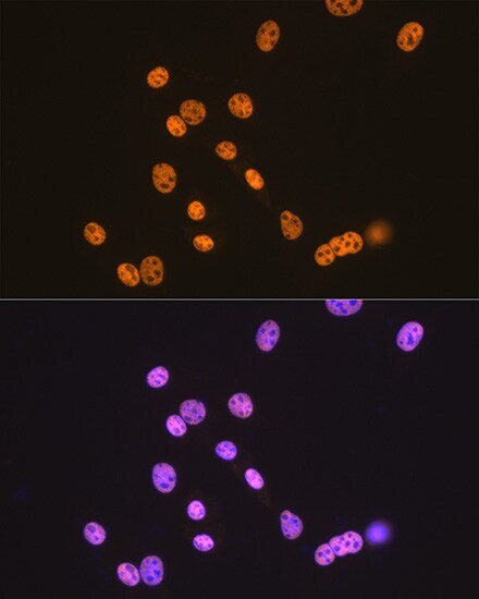
- Immunocytochemical analysis of TDP-43 performed on NIH-3T3 cells using a TDP-43 monoclonal antibody (Product # MA5-35273) at a dilution of 1:100. An HRP Goat Anti-Rabbit IgG (H+L) secondary antibody was applied at a dilution of 1:10,000.
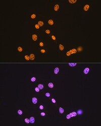
- Submitted by
- Invitrogen Antibodies (provider)
- Main image
- Experimental details
- Immunocytochemical analysis of TDP-43 performed on C6 cells using a TDP-43 monoclonal antibody (Product # MA5-35273) at a dilution of 1:100. An HRP Goat Anti-Rabbit IgG (H+L) secondary antibody was applied at a dilution of 1:10,000.
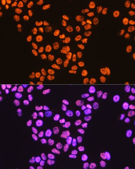
- Submitted by
- Invitrogen Antibodies (provider)
- Main image

- Experimental details
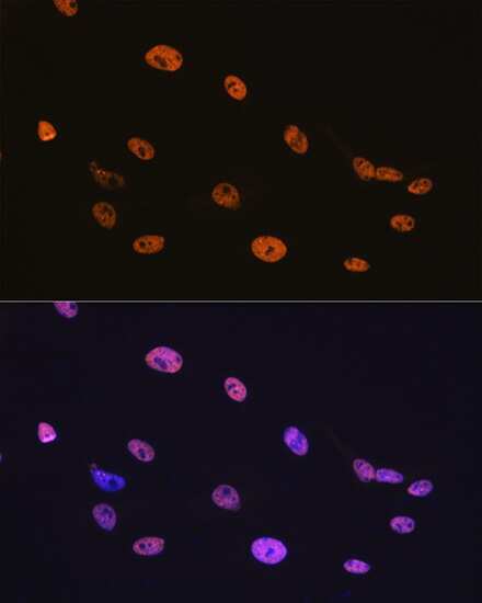
- Immunocytochemical analysis of TDP-43 performed on HeLa cells using a TDP-43 monoclonal antibody (Product # MA5-35273) at a dilution of 1:100. An HRP Goat Anti-Rabbit IgG (H+L) secondary antibody was applied at a dilution of 1:10,000.
- Submitted by
- Invitrogen Antibodies (provider)
- Main image
- Experimental details
- Immunocytochemical analysis of TDP-43 performed on NIH-3T3 cells using a TDP-43 monoclonal antibody (Product # MA5-35273) at a dilution of 1:100. An HRP Goat Anti-Rabbit IgG (H+L) secondary antibody was applied at a dilution of 1:10,000.
- Submitted by
- Invitrogen Antibodies (provider)
- Main image

- Experimental details
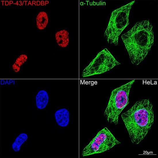
- Confocal imaging (Immunocytochemistry) of TDP-43 in HeLa cells. Samples were incubated with TDP-43 Monoclonal antibody (Product # MA5-35273) using a dilution of 1:100 (Red). The cells were counterstained with alpha Tubulin Mouse mAb (dilution 1:400) (Green). DAPI was used for nuclear staining (blue). Objective: 100x.
Supportive validation
- Submitted by
- Invitrogen Antibodies (provider)
- Main image
- Experimental details
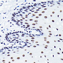
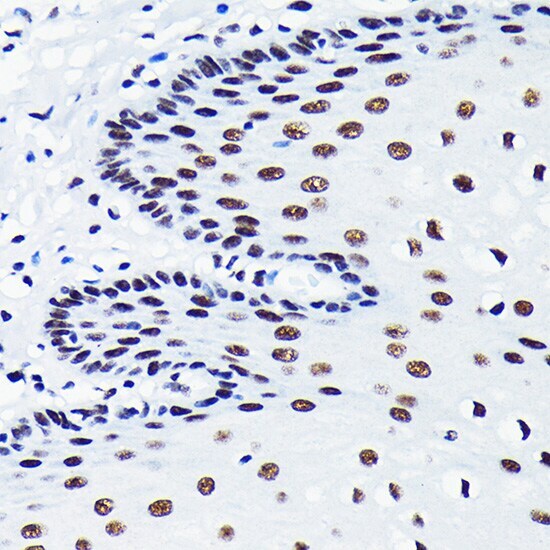
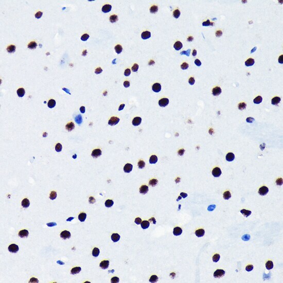
- Immunohistochemistry analysis of TDP-43 in paraffin-embedded human esophageal. Samples were incubated with TDP-43 Monoclonal antibody (Product # MA5-35273) using a dilution of 1:100 (40x lens). Perform microwave antigen retrieval with 10 mM PBS buffer pH 7.2 before commencing with IHC staining protocol.
- Submitted by
- Invitrogen Antibodies (provider)
- Main image
- Experimental details
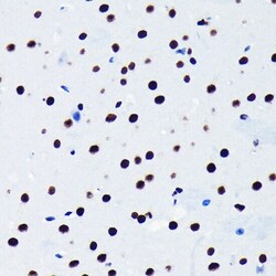
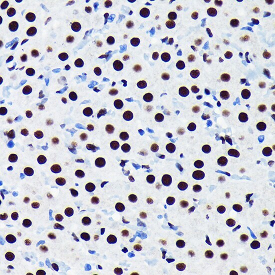
- Immunohistochemistry analysis of TDP-43 in paraffin-embedded mouse brain. Samples were incubated with TDP-43 Monoclonal antibody (Product # MA5-35273) using a dilution of 1:100 (40x lens). Perform microwave antigen retrieval with 10 mM PBS buffer pH 7.2 before commencing with IHC staining protocol.
- Submitted by
- Invitrogen Antibodies (provider)
- Main image
- Experimental details
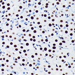
- Immunohistochemistry analysis of TDP-43 in paraffin-embedded rat ovary. Samples were incubated with TDP-43 Monoclonal antibody (Product # MA5-35273) using a dilution of 1:100 (40x lens). Perform microwave antigen retrieval with 10 mM PBS buffer pH 7.2 before commencing with IHC staining protocol.
Supportive validation
- Submitted by
- Invitrogen Antibodies (provider)
- Main image
- Experimental details
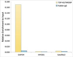
- ChIP analysis of TDP-43 in extracts of K562 cells. Samples were incubated with TDP-43 Monoclonal antibody (Product # MA5-35273) and rabbit IgG. The amount of immunoprecipitated DNA was checked by quantitative PCR. Histogram was constructed by the ratios of the immunoprecipitated DNA to the input.

Supportive validation
- Submitted by
- Invitrogen Antibodies (provider)
- Main image
- Experimental details
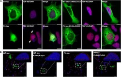
- FIGURE 5 PH-tau-positive cells present mislocalization of TDP-43 to the cytoplasm when PH-tau can enter the nucleus, but not when it is kept in the cytoplasm. (A) HEK-293 cells were transfected with WT-tau, PH-tau, WT-tau-K140A,K141A, or PH-tau-K140A,K141A (green) and immunostained for TDP-43 (red). The localization of TDP-43 was determined by looking for co-localization of the TDP-43 stain and DAPI (blue), which presents as purple. The arrows indicate areas of red staining that do not co-localize with DAPI, indicating mislocalization to the cytoplasm. Scale bar indicates 15 mum. (B) Overlays of z-stack images taken with a confocal microscope. The inset displays an IMARIS reconstruction of the indicated cell to better display the nuclear or cytoplasmic localization of TDP-43 (red dots). Scale bar indicates 15 mum.
