Explore
 Validate
 Learn
Learn10782-2-AP
antibody from Invitrogen Antibodies
Targeting: TARDBP
ALS10, TDP-43
 Western blot Immunocytochemistry
 Immunoprecipitation Immunohistochemistry Flow cytometry
 Chromatin Immunoprecipitation Other assay
Antibody data
- Antibody Data
- Antigen structure
- References [0]
- Comments [0]
- Validations
- Western blot [8]
- Immunocytochemistry [5]
- Immunohistochemistry [14]
- Flow cytometry [1]
- Other assay [2]
Submit
Validation data
Reference
Comment
Report error
- Product number
- 10782-2-AP - Provider product page

- Provider
- Invitrogen Antibodies
- Product name
- TDP-43 Polyclonal Antibody
- Antibody type
- Polyclonal
- Antigen
- Other
- Reactivity
- Human, Mouse, Rat, Zebrafish
- Host
- Rabbit
- Isotype
- IgG
- Vial size
- 150 µL
- Concentration
- 0.31 mg/mL
- Storage
- -20°C
No comments: Submit comment
Supportive validation
- Submitted by
- Invitrogen Antibodies (provider)
- Main image

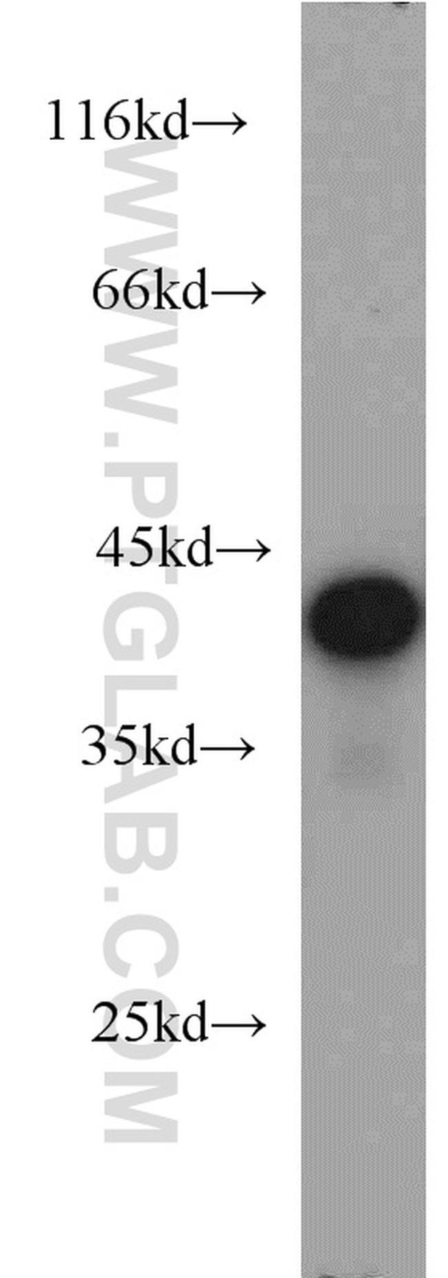
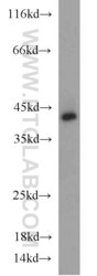
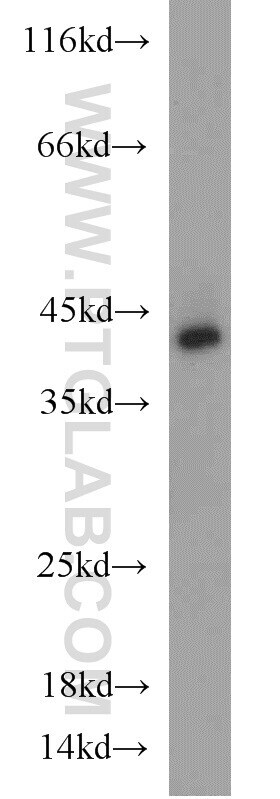
- Experimental details
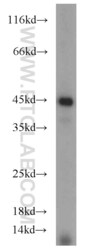
- HeLa cells were subjected to SDS PAGE followed by western blot with 10782-2-AP (TARDBP antibody) at dilution of 1:1000 incubated at room temperature for 1.5 hours.
- Submitted by
- Invitrogen Antibodies (provider)
- Main image
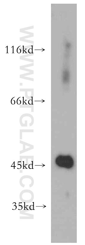
- Experimental details
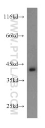
- Mouse pancreas tissue were subjected to SDS PAGE followed by western blot with 10782-2-AP (TARDBP antibody) at dilution of 1:800 incubated at room temperature for 1.5 hours.
- Submitted by
- Invitrogen Antibodies (provider)
- Main image
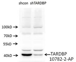
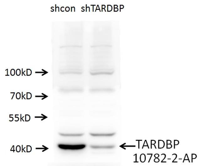
- Experimental details
- A549 cells (shcontrol and shRNA of TDP43) were subjected to SDS PAGE followed by western blot with 10782-2-AP (TARDBP antibody) at dilution of 1:1000.
- Submitted by
- Invitrogen Antibodies (provider)
- Main image
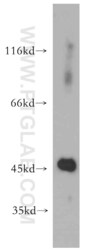
- Experimental details
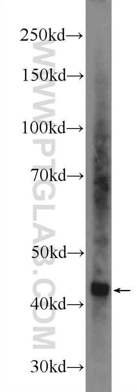
- Human heart tissue were subjected to SDS PAGE followed by western blot with 10782-2-AP (TARDBP antibody) at dilution of 1:500 incubated at room temperature for 1.5 hours.
- Submitted by
- Invitrogen Antibodies (provider)
- Main image
- Experimental details
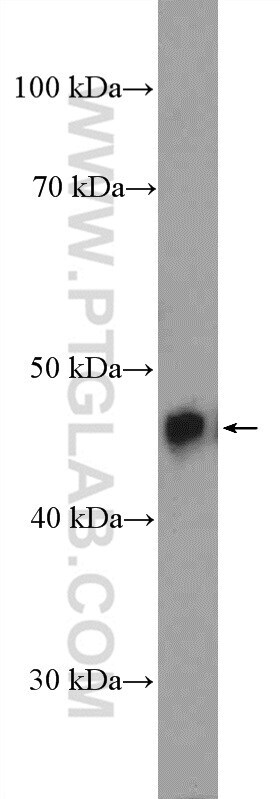
- HL-60 cells were subjected to SDS PAGE followed by western blot with 10782-2-AP (TARDBP antibody) at dilution of 1:500 incubated at room temperature for 1.5 hours.

- Submitted by
- Invitrogen Antibodies (provider)
- Main image
- Experimental details
- K-562 cells were subjected to SDS PAGE followed by western blot with 10782-2-AP (TARDBP antibody) at dilution of 1:6000 incubated at room temperature for 1.5 hours.

- Submitted by
- Invitrogen Antibodies (provider)
- Main image

- Experimental details
- Mouse brain tissue were subjected to SDS PAGE followed by western blot with 10782-2-AP (TDP-43 Antibody) at dilution of 1:600 incubated at room temperature for 1.5 hours.
- Submitted by
- Invitrogen Antibodies (provider)
- Main image

- Experimental details
- Rat brain tissue were subjected to SDS PAGE followed by western blot with 10782-2-AP (TDP-43 Antibody) at dilution of 1:600 incubated at room temperature for 1.5 hours.
Supportive validation
- Submitted by
- Invitrogen Antibodies (provider)
- Main image
- Experimental details
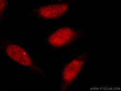
- Immunofluorescent analysis of MCF-7 cells, using TARDBP antibody 10782-2-AP at 1:50 dilution and Rhodamine-labeled goat anti-rabbit IgG (red).

- Submitted by
- Invitrogen Antibodies (provider)
- Main image
- Experimental details
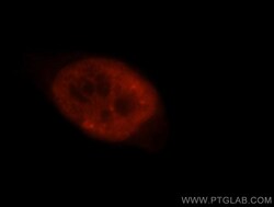
- Immunofluorescent analysis of SH-SY5Y cells using 10782-2-AP (TDP-43 Antibody) at dilution of 1:50 and Alexa Fluor 488-conjugated AffiniPure Goat Anti-Rabbit IgG (H+L). Cells were fixed with 10% Formaldehyde.

- Submitted by
- Invitrogen Antibodies (provider)
- Main image

- Experimental details
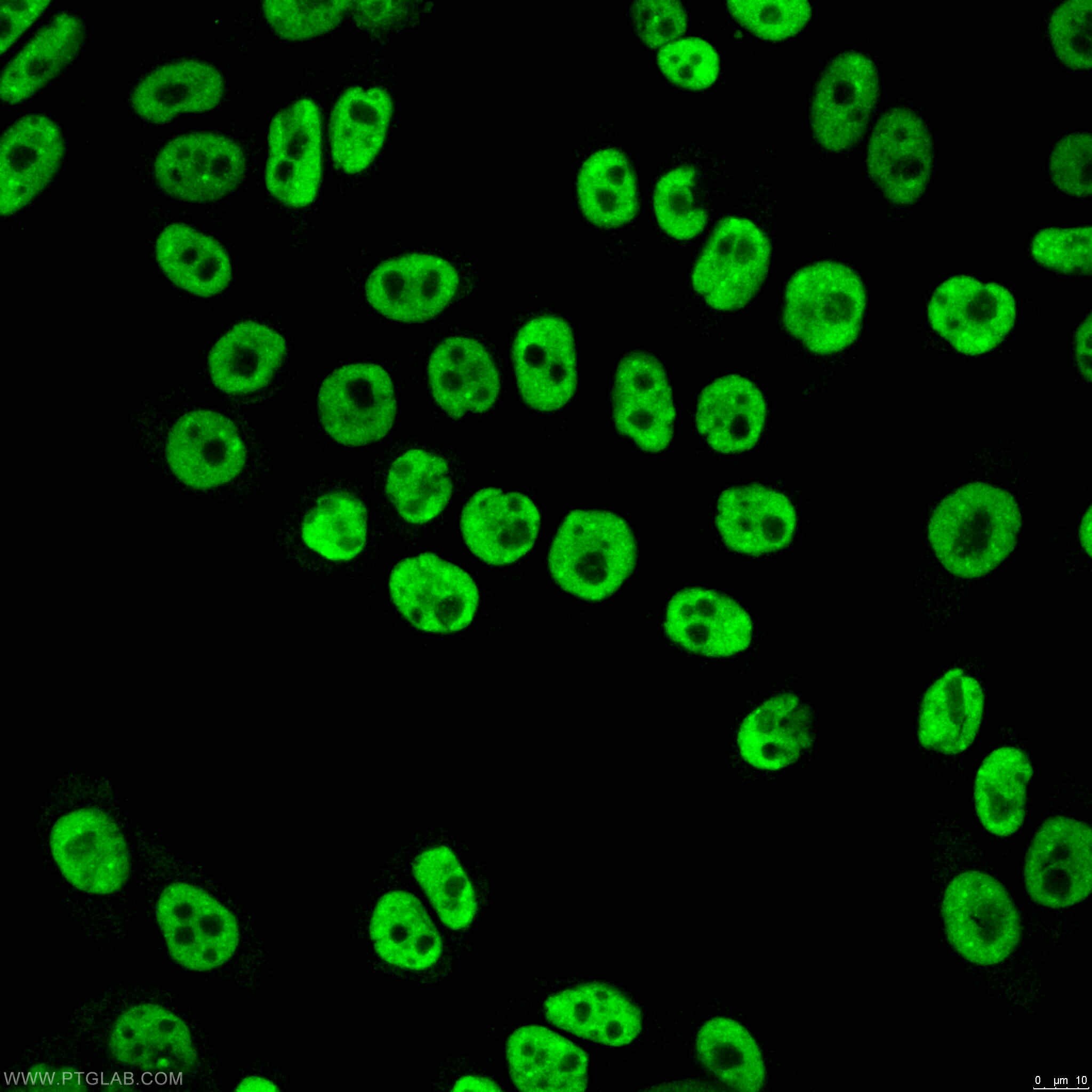
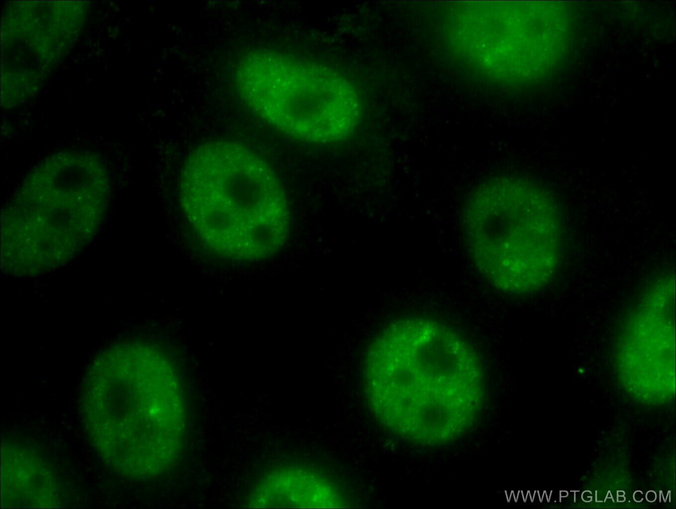
- Immunofluorescent analysis of ( 4% PFA) fixed HeLa cells using 10782-2-AP (TDP-43 antibody) at dilution of 1:100 and Alexa Fluor 488-Conjugated AffiniPure Goat Anti-Rabbit IGG (H+L).
- Submitted by
- Invitrogen Antibodies (provider)
- Main image

- Experimental details
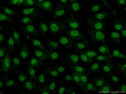
- Immunofluorescent analysis of (10% Formaldehyde) fixed HeLa cells using 10782-2-AP (TDP-43 antibody) at dilution of 1:400 and Alexa Fluor 488-conjugated AffiniPure Goat Anti-Rabbit IgG (H+L).
- Submitted by
- Invitrogen Antibodies (provider)
- Main image
- Experimental details
- Immunofluorescent analysis of HepG2 cells, using TARDBP antibody 10782-2-AP at 1:50 dilution and Rhodamine-labeled goat anti-rabbit IgG (red).

Supportive validation
- Submitted by
- Invitrogen Antibodies (provider)
- Main image
- Experimental details
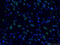
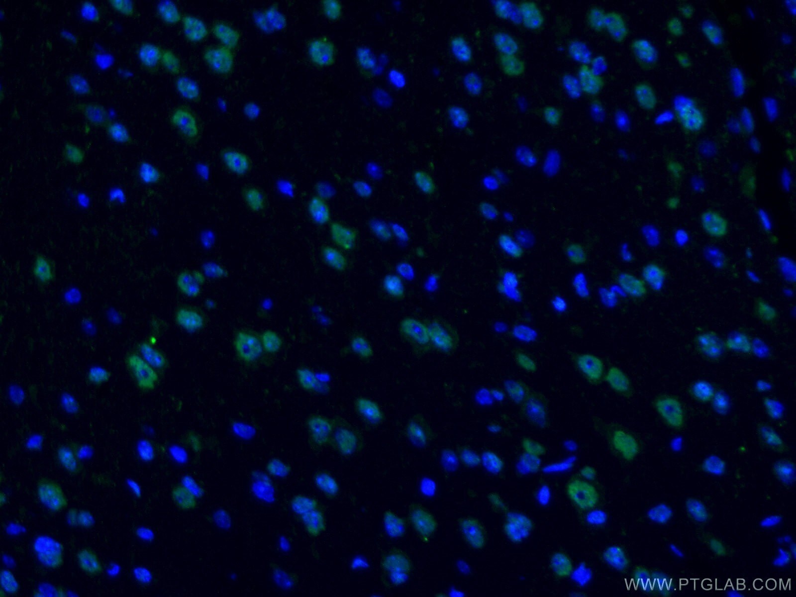
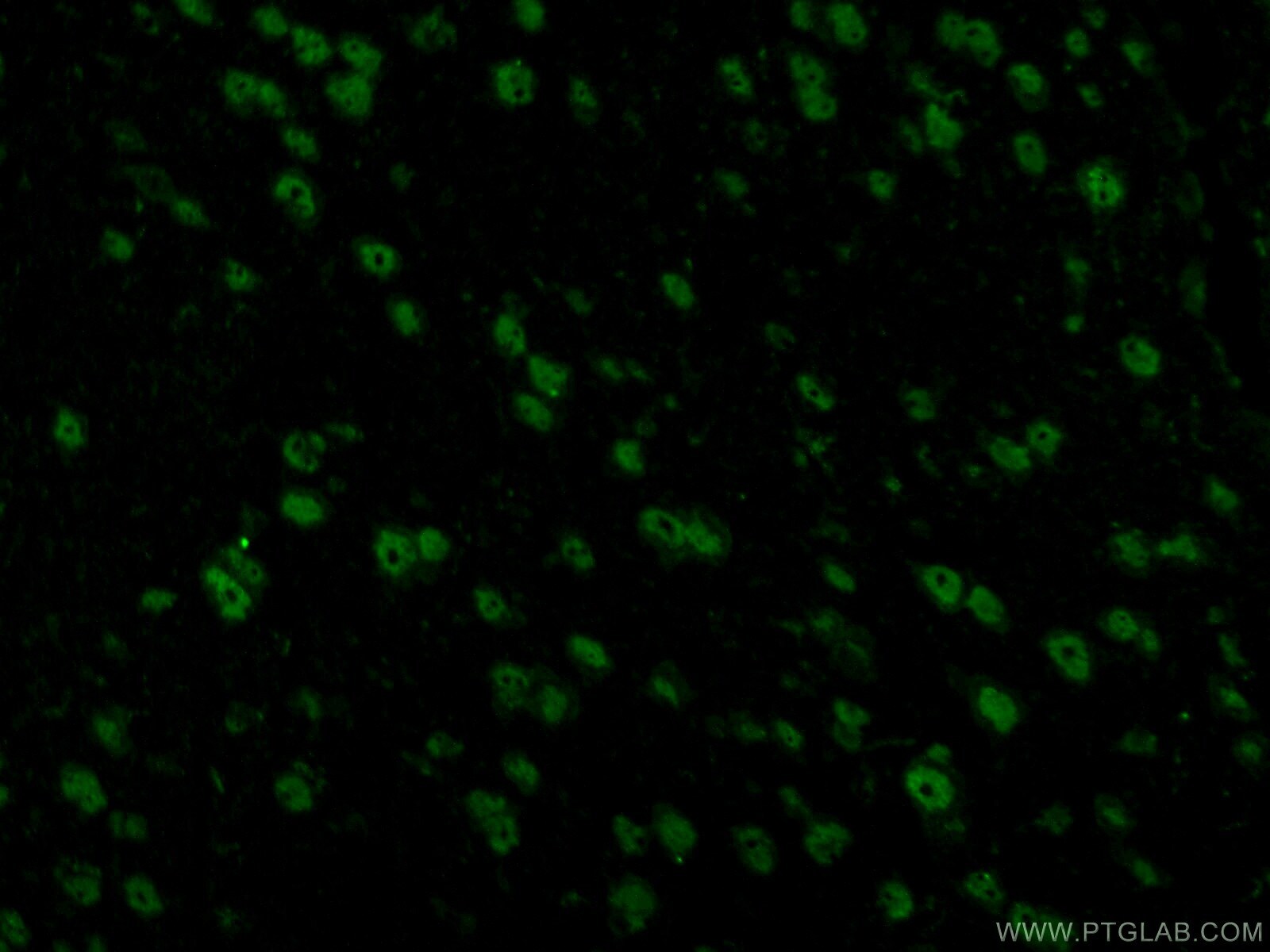
- Immunofluorescent analysis of (4% PFA) fixed mouse brain tissue using 10782-2-AP (TDP-43 antibody) at dilution of 1:50 and Alexa Fluor 488-conjugated AffiniPure Goat Anti-Rabbit IgG (H+L).
- Submitted by
- Invitrogen Antibodies (provider)
- Main image

- Experimental details
- Immunofluorescent analysis of (4% PFA) fixed mouse brain tissue using 10782-2-AP (TDP-43 antibody) at dilution of 1:50 and Alexa Fluor 488-conjugated AffiniPure Goat Anti-Rabbit IgG (H+L).
- Submitted by
- Invitrogen Antibodies (provider)
- Main image
- Experimental details
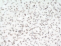
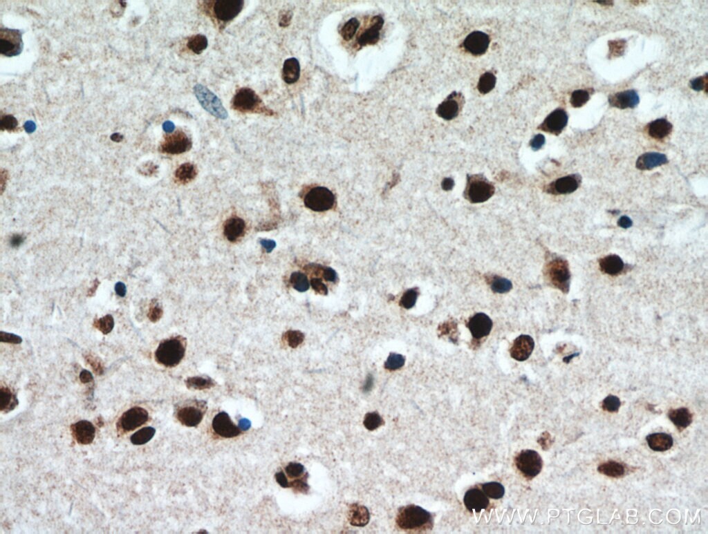
- Immunohistochemistry of paraffin-embedded human brain using 10782-2-AP (TARDBP antibody) at dilution of 1:50 (under 10x lens).
- Submitted by
- Invitrogen Antibodies (provider)
- Main image

- Experimental details
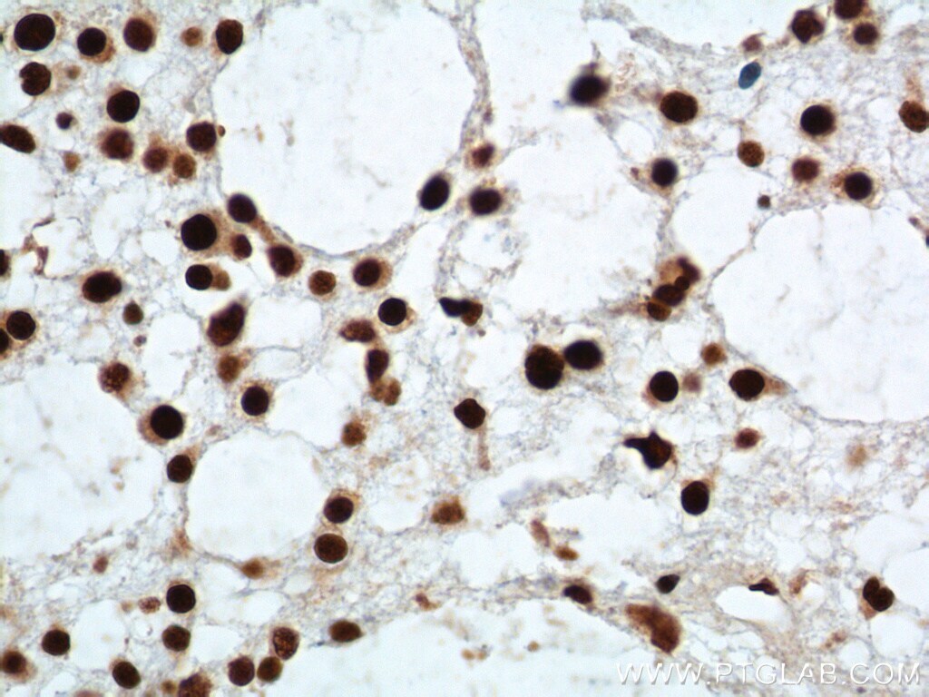
- Immunohistochemistry of paraffin-embedded human gliomas tissue slide using 10782-2-AP (TDP-43 Antibody) at dilution of 1:2000 (under 40x lens). heat mediated antigen retrieved with Tris-EDTA buffer (pH 9).
- Submitted by
- Invitrogen Antibodies (provider)
- Main image
- Experimental details
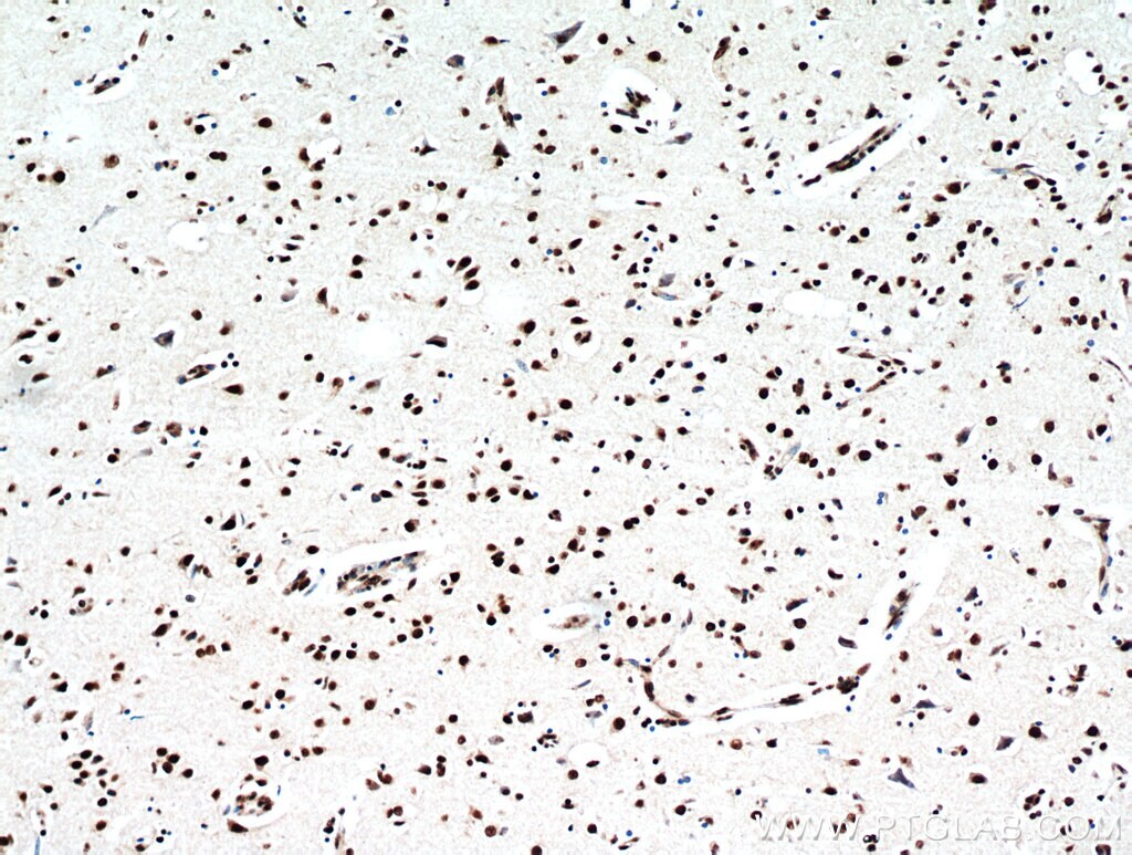
- Immunohistochemistry of paraffin-embedded human brain using 10782-2-AP (TARDBP antibody) at dilution of 1:50 (under 40x lens).
- Submitted by
- Invitrogen Antibodies (provider)
- Main image

- Experimental details
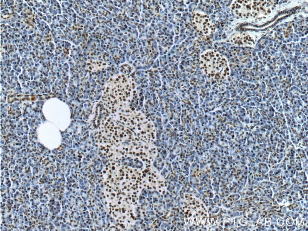
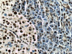
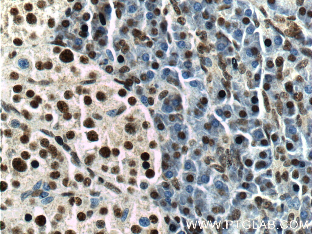
- Immunohistochemistry of paraffin-embedded human pancreas tissue slide using 10782-2-AP (TDP-43 Antibody) at dilution of 1:200 (under 10x lens).
- Submitted by
- Invitrogen Antibodies (provider)
- Main image
- Experimental details
- Immunohistochemistry of paraffin-embedded human pancreas tissue slide using 10782-2-AP (TDP-43 Antibody) at dilution of 1:200 (under 40x lens).
- Submitted by
- Invitrogen Antibodies (provider)
- Main image
- Experimental details
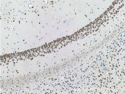
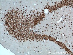
- Immunohistochemistry of paraffin-embedded mouse brain tissue slide using 10782-2-AP (TDP-43 Antibody) at dilution of 1:200 (under 10x lens).
- Submitted by
- Invitrogen Antibodies (provider)
- Main image
- Experimental details
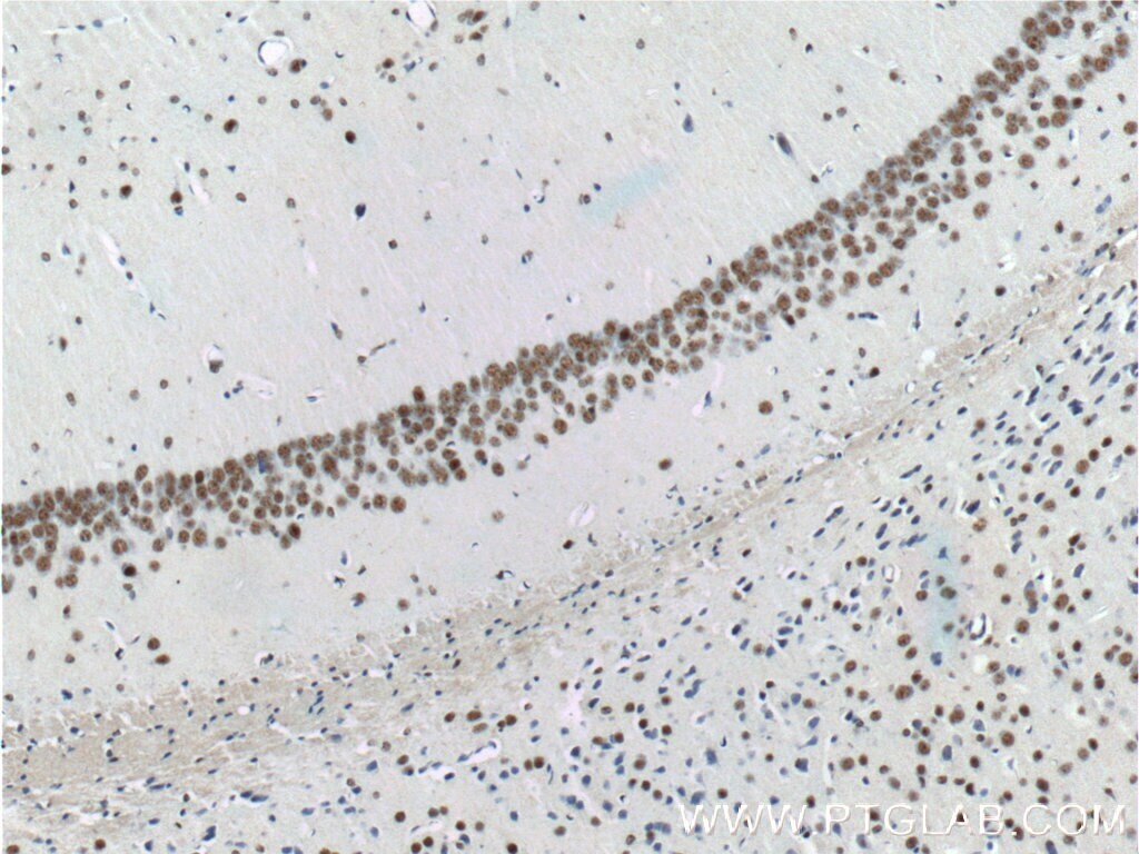
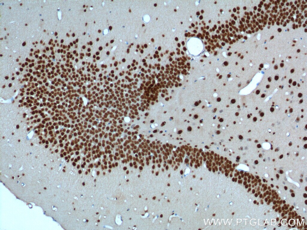
- Immunohistochemistry of paraffin-embedded mouse brain tissue slide using 10782-2-AP (TDP-43 Antibody) at dilution of 1:200 (under 40x lens).
- Submitted by
- Invitrogen Antibodies (provider)
- Main image
- Experimental details
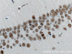
- Immunohistochemistry of paraffin-embedded rat brain tissue slide using 10782-2-AP (TDP-43 Antibody) at dilution of 1:200 (under 10x lens).
- Submitted by
- Invitrogen Antibodies (provider)
- Main image

- Experimental details
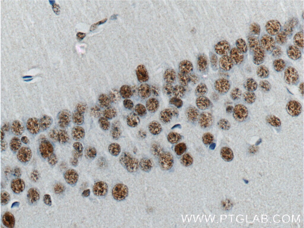
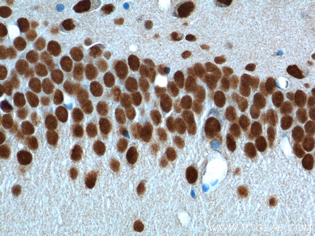
- Immunohistochemistry of paraffin-embedded rat brain tissue slide using 10782-2-AP (TDP-43 Antibody) at dilution of 1:200 (under 40x lens).
- Submitted by
- Invitrogen Antibodies (provider)
- Main image
- Experimental details
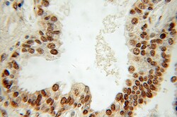
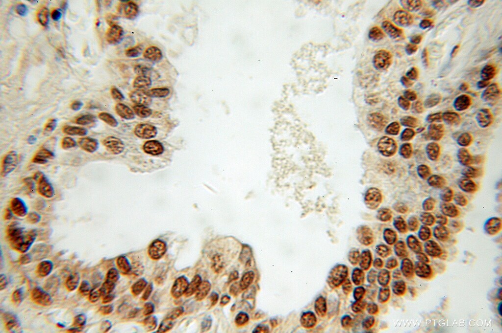
- Immunohistochemistry of paraffin-embedded human prostate cancer using 10782-2-AP (TARDBP antibody) at dilution of 1:100 (under 10x lens).
- Submitted by
- Invitrogen Antibodies (provider)
- Main image
- Experimental details
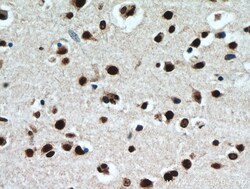
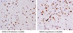
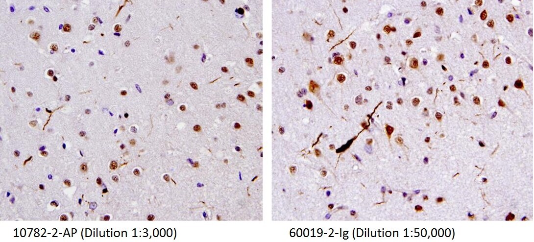
- 40X of FTLD-U case stained by 10782-2-AP and 60019-2-IG, showing dystrophic neurites. (Figs were provided by Linda K. Kwong).
- Submitted by
- Invitrogen Antibodies (provider)
- Main image

- Experimental details
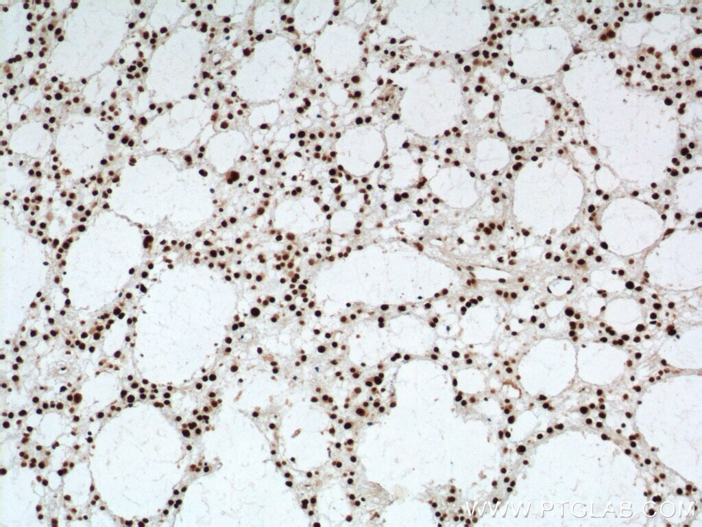
- Immunohistochemistry of paraffin-embedded human gliomas tissue slide using 10782-2-AP ( TDP-43 Antibody) at dilution of 1:2000 (under 10x lens). heat mediated antigen retrieved with Tris-EDTA buffer (pH 9).
Supportive validation
- Submitted by
- Invitrogen Antibodies (provider)
- Main image
- Experimental details
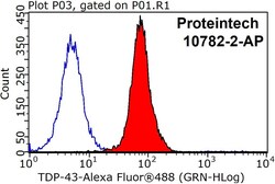
- 1X10^6 HeLa cells were stained with 0.2ug TDP-43 antibody (10782-2-AP, red) and control antibody (blue). Fixed with 90% MeOH blocked with 3% BSA (30 min). Alexa Fluor 488-conjugated AffiniPure Goat Anti-Rabbit IGG (H+L) with dilution 1:1000.

Supportive validation
- Submitted by
- Invitrogen Antibodies (provider)
- Main image

- Experimental details
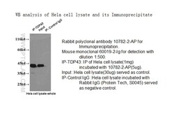
- IP result of anti-TDP-43 (IP:10782-2-AP, 3ug; Detection:10782-2-AP 1:1000) with K-562 cells lysate 1000ug.
- Submitted by
- Invitrogen Antibodies (provider)
- Main image
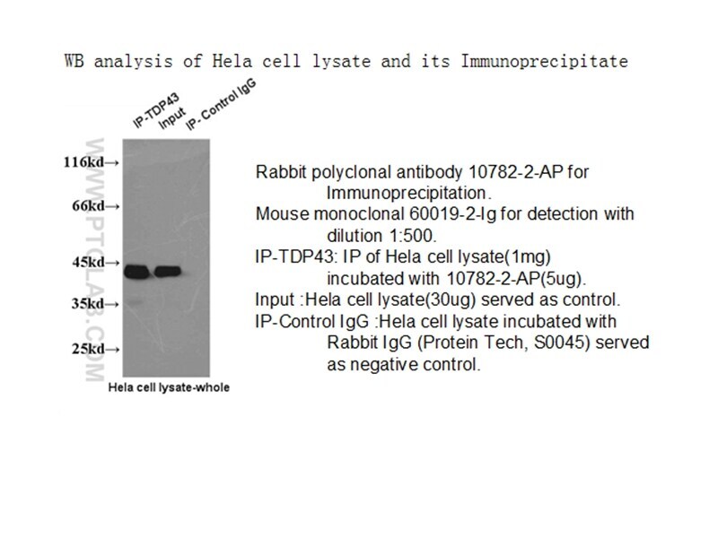
- Experimental details
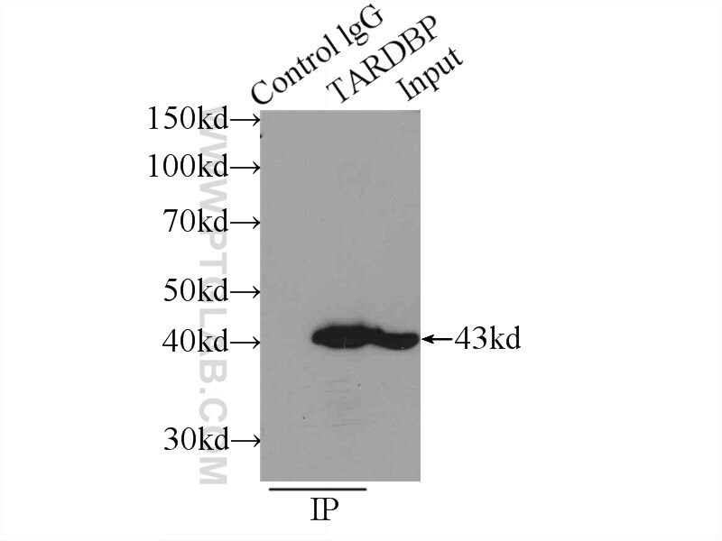
- IP result of anti-TDP43 (10782-2-AP for IP and 60019-2-Ig for Detection).