 Explore
 Validate
 Learn
Learn Western blot
 Immunoprecipitation
Antibody data
- Antibody Data
- Antigen structure
- References [2]
- Comments [0]
- Validations
- Western blot [2]
- Immunocytochemistry [1]
- Other assay [1]
Submit
Validation data
Reference
Comment
Report error
- Product number
- PA5-17011 - Provider product page

- Provider
- Invitrogen Antibodies
- Product name
- TDP-43 Polyclonal Antibody
- Antibody type
- Polyclonal
- Antigen
- Synthetic peptide
- Description
- It is not recommended to aliquot this antibody.
- Reactivity
- Human, Mouse, Rat
- Host
- Rabbit
- Isotype
- IgG
- Vial size
- 100 µL
- Concentration
- 14 µg/mL
- Storage
- -20°C
Submitted references Muscle cells of sporadic amyotrophic lateral sclerosis patients secrete neurotoxic vesicles.
Autophagy induction enhances TDP43 turnover and survival in neuronal ALS models.
Le Gall L, Duddy WJ, Martinat C, Mariot V, Connolly O, Milla V, Anakor E, Ouandaogo ZG, Millecamps S, Lainé J, Vijayakumar UG, Knoblach S, Raoul C, Lucas O, Loeffler JP, Bede P, Behin A, Blasco H, Bruneteau G, Del Mar Amador M, Devos D, Henriques A, Hesters A, Lacomblez L, Laforet P, Langlet T, Leblanc P, Le Forestier N, Maisonobe T, Meininger V, Robelin L, Salachas F, Stojkovic T, Querin G, Dumonceaux J, Butler Browne G, González De Aguilar JL, Duguez S, Pradat PF
Journal of cachexia, sarcopenia and muscle 2022 Apr;13(2):1385-1402
Journal of cachexia, sarcopenia and muscle 2022 Apr;13(2):1385-1402
Autophagy induction enhances TDP43 turnover and survival in neuronal ALS models.
Barmada SJ, Serio A, Arjun A, Bilican B, Daub A, Ando DM, Tsvetkov A, Pleiss M, Li X, Peisach D, Shaw C, Chandran S, Finkbeiner S
Nature chemical biology 2014 Aug;10(8):677-85
Nature chemical biology 2014 Aug;10(8):677-85
No comments: Submit comment
Supportive validation
- Submitted by
- Invitrogen Antibodies (provider)
- Main image
- Experimental details
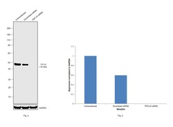
- Knockdown of TDP-43 was achieved by transfecting HeLa with TDP-43 specific siRNAs (Silencer® select Product # s23830, s23829). Western blot analysis (Fig. a) was performed using Whole Cell Extract-WCL from the TDP-43 knockdown cells (lane 3), non-targeting scrambled siRNA transfected cells (lane 2) and untransfected cells (lane 1). The blot was probed with TDP-43 Polyclonal Antibody (Product # PA5-17011, 1:1000 dilution) and Goat anti-Rabbit IgG (H+L) Superclonal™ Recombinant Secondary Antibody, HRP (Product # A27036, 1:4000 dilution). Densitometric analysis of this western blot is shown in histogram (Fig. b). Decrease in signal upon siRNA mediated knock down confirms that antibody is specific to TDP-43.

- Submitted by
- Invitrogen Antibodies (provider)
- Main image

- Experimental details
- Western blot was performed using Anti-TDP-43 Polyclonal Antibody(Product # PA5-17011) and a 44kDa band corresponding to TDP-43 was observed across cell lines and tissue extract tested. A non-specific band around 80 kDa has also been observed. Whole Cell Extract-WCL (30 µg lysate) of HeLa (Lane 1), MCF7 (Lane 2), Caco-2 (Lane 3), K-562 (Lane 4), Neuro-2a (Lane 5), NIH/3T3 (Lane 6) and tissue extract of Mouse Thymus (Lane 7), were electrophoresed using NuPAGE™ 4-12% Bis-Tris Protein Gel (Product # NP0322BOX). Resolved proteins were then transferred onto a Nitrocellulose membrane (Product # IB23001) by iBlot® 2 Dry Blotting System (Product # IB21001). The blot was probed with the primary antibody (1:1000 dilution) and detected by chemiluminescence with Goat anti-Rabbit IgG (H+L) Superclonal™ Recombinant Secondary Antibody, HRP (Product # A27036, 1:4000 dilution) using the iBright FL 1000 (Product # A32752). Chemiluminescent detection was performed using Novex® ECL Chemiluminescent Substrate Reagent Kit (Product # WP20005).

Supportive validation
- Submitted by
- Invitrogen Antibodies (provider)
- Main image
- Experimental details
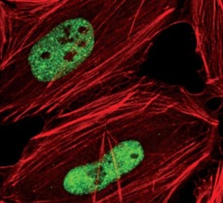
- Immunofluorescent analysis of TDP43 in HeLa cells using a TDP43 polyclonal antibody (Product # PA5-17011) (green). Actin filaments are labeled with a fluorescent red phalloidin.

Supportive validation
- Submitted by
- Invitrogen Antibodies (provider)
- Main image

- Experimental details
- Figure 2 TDP43 turnover in primary neurons (a) Primary neurons were labeled with 35 S-labeled methionine, and endogenous TDP43 was immunoprecipitated using anti-TDP43 antibodies (IP-TDP43) or non-specific IgG antibodies (IP-IgG). (b) Log-normal plot of normalized TDP43 levels. TDP43 half-life was determined by fitting a first-order exponential curve to the data (R 2 = 0.8984). Values pooled from five independent experiments. (c) Potential confounders in half-life analyses. Ideally (top), total protein decreases over time in a predictable manner (top right panel). Toxicity (middle) reduces the amount of measured protein, potentially shortening the calculated half-life (middle right panel). Protein aggregation (bottom) might also shorten half-life estimations if the protein cannot be immunoprecipitated (dark arrows, bottom right panel), or lengthen it if the protein is more stable and successfully immunoprecipitated (grey arrows, bottom right panel). (d) Optical pulse labeling. Primary cortical neurons were transfected with EGFP and TDP43(WT)-Dendra2, photoconverted then imaged by AFM. Dead cells (arrow) were excluded at the time of death. (e) The half-life of TDP43(WT)-Dendra2 was determined by fitting a first-order exponential curve to the data (R 2 0.9558). (f) Including cells with aggregates prolonged TDP43(WT)-Dendra2 half-life (purple line, R 2 = 0.9003; * p < 0.0001, F 15.34, extra sum-of-squares F-test). Excluding cells that died over the 36 h experiment also prolonged
