Explore
 Validate
 Learn
Learn Western blot
Antibody data
- Antibody Data
- Antigen structure
- References [0]
- Comments [0]
- Validations
- Western blot [3]
- Immunocytochemistry [1]
Submit
Validation data
Reference
Comment
Report error
- Product number
- PA5-19712 - Provider product page

- Provider
- Invitrogen Antibodies
- Product name
- TDP-43 Polyclonal Antibody
- Antibody type
- Polyclonal
- Antigen
- Synthetic peptide
- Description
- PA5-19712 targets TARDBP in IF, IHC (F), IP, and WB applications and shows reactivity with Human and mouse samples.
- Concentration
- 0.5 mg/mL
No comments: Submit comment
Supportive validation
- Submitted by
- Invitrogen Antibodies (provider)
- Main image
- Experimental details
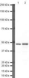
- Western blot analysis of HepG2 Nuclear Lysate using Product # PA5-19712, TARDBP primary antibody at a dilution of 1 µg/mL (lane 1). Staining of HeLa Nuclear Lysate at a dilution of 1 µg/mL (lane 2). Blot treated with a secondary HRP-conjugated Goat polyclonal anti-Rabbit antibody was used at a dilution of 1:3000.

- Submitted by
- Invitrogen Antibodies (provider)
- Main image
- Experimental details
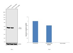
- Knockdown of TDP-43 was achieved by transfecting HeLa with TDP-43 specific siRNAs (Silencer® select Product # s23830, s23829). Western blot analysis (Fig. a) was performed using Whole Cell Extract-WCL from the TDP-43 knockdown cells (lane 3), non-targeting scrambled siRNA transfected cells (lane 2) and untransfected cells (lane 1). The blot was probed with TDP-43 Polyclonal Antibody (Product # PA5-19712, 1 ug/ml ) and Goat anti-Rabbit IgG (H+L) Superclonal™ Recombinant Secondary Antibody, HRP (Product # A27036, 1:4000 dilution). Densitometric analysis of this western blot is shown in histogram (Fig. b). Decrease in signal upon siRNA mediated knock down confirms that antibody is specific to TDP-43.

- Submitted by
- Invitrogen Antibodies (provider)
- Main image

- Experimental details
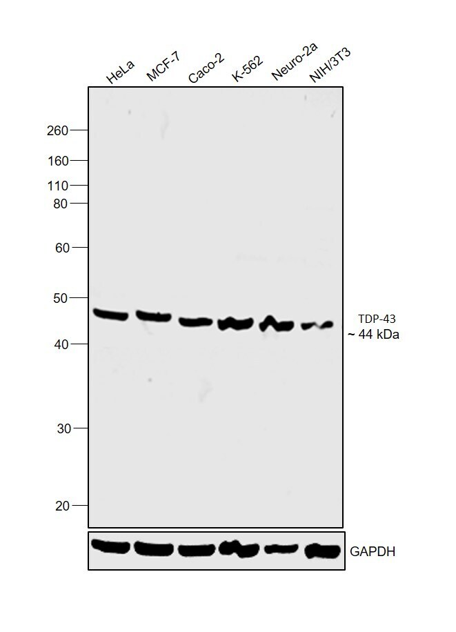
- Western blot was performed using Anti-TDP-43 Polyclonal Antibody(Product # PA5-19712) and a 44kDa band corresponding to TDP-43 was observed across cell lines tested. Whole Cell Extract-WCL (30 µg lysate) of HeLa (Lane 1), MCF7 (Lane 2), Caco-2 (Lane 3), K-562 (Lane 4), Neuro-2a (Lane 5) and NIH/3T3 (Lane 6) were electrophoresed using NuPAGE™ 4-12% Bis-Tris Protein Gel (Product # NP0322BOX). Resolved proteins were then transferred onto a Nitrocellulose membrane (Product # IB23001) by iBlot® 2 Dry Blotting System (Product # IB21001). The blot was probed with the primary antibody (1 µg/mL) and detected by chemiluminescence with Goat anti-Rabbit IgG (H+L) Superclonal™ Recombinant Secondary Antibody, HRP (Product # A27036,1:4000 dilution) using the iBright FL 1000 (Product # A32752). Chemiluminescent detection was performed using Novex® ECL Chemiluminescent Substrate Reagent Kit (Product # WP20005).
Supportive validation
- Submitted by
- Invitrogen Antibodies (provider)
- Main image
- Experimental details
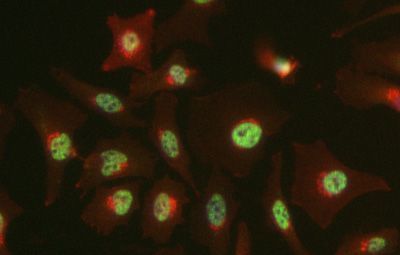
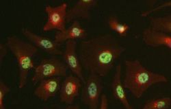
- Immunofluorescent staining of Hek293 cells using Product # PA5-19712, anti-TARDBP antibody. The cells were fixed with PFA (4%) for 10 minutes, permabilised with BSA (1%), normal goat serum (10%) and glycine (0.3 M) in 0.1% T-BST for 1 hour and exposed to the primary antibody at a concentration of 5 µg/mL for 1 hour at room temp. The secondary antibody was a 448 fluorescence conjugated Goat anti-rabbit IgG (green) at a dilution of 1:1000. A WGA- 594 fluorescent conjugated stain was used to label plasma membranes (red) and the nuclei stain was DAPI (blue).